021 202 0626
What to Expect During a Gastroscopy Procedure: Step-by-Step Guide

What to Expect During a Gastroscopy Procedure: Step-by-Step Guide

Introduction


For individuals facing gastrointestinal issues, the prospect of undergoing a gastroscopy procedure can be both daunting and mysterious.

Gastroscopy, also known as an upper endoscopy, is a medical procedure used to examine the upper digestive tract, providing crucial insights into potential issues within the esophagus, stomach, and the first part of the small intestine.

In this comprehensive guide, we will walk you through what to expect during a gastroscopy procedure, step by step, alleviating uncertainties and empowering you with knowledge.


What is Gastroscopy?


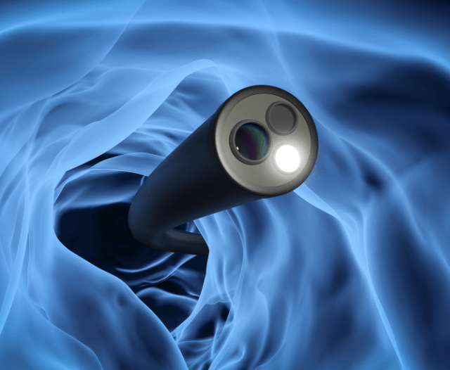
Gastroscopy involves the use of a thin, flexible tube called an endoscope, equipped with a light and a camera, to visualize and capture images of the digestive organs.

This procedure is commonly employed to diagnose and investigate various gastrointestinal conditions, including gastroesophageal reflux disease (GERD), ulcers, inflammation, and even to screen for certain cancers.

Gastroesophageal reflux disease:
Gastroesophageal reflux disease (GERD) is a chronic digestive disorder characterized by the reflux of stomach acid into the esophagus, causing symptoms such as heartburn, regurgitation, chest pain, and difficulty swallowing.

It occurs when the lower esophageal sphincter (LES)—a muscular ring at the junction between the esophagus and the stomach—does not function properly, allowing stomach contents to flow back up into the esophagus.

Ulcer:  An ulcer is a type of open sore or lesion that forms on the skin or mucous membranes of the body.

When discussing ulcers in the context of gastrointestinal health, we typically refer to peptic ulcers, which occur in the lining of the stomach, esophagus, or duodenum (the first part of the small intestine).


Gastrointestinal inflammation:
Gastrointestinal inflammation refers to a condition characterized by irritation, swelling, and damage to the lining of the gastrointestinal tract.

This inflammation can affect various parts of the digestive system, including the esophagus, stomach, small intestine, and large intestine.

It can be caused by a wide range of factors, including infections, autoimmune disorders, dietary factors, and medications.


Before the Procedure


Preparation Instructions:

Before the gastroscopy, your healthcare provider will provide detailed instructions on how to prepare for the procedure.

This may include fasting for a specific period, typically overnight, to ensure a clear view of the digestive tract.


Medication Adjustments:

Inform your healthcare provider about any medications you are currently taking, especially blood thinners or anticoagulants.

In some cases, adjustments to your medication regimen may be necessary before the procedure.


Health Assessment:

A pre-procedure health assessment will be conducted to ensure that you are physically prepared for the gastroscopy.

This may involve checking vital signs, reviewing your medical history, and addressing any concerns you may have.

gastroscopy_procedure

The Gastroscopy Procedure


Arrival at the Medical Facility:

On the day of the procedure, you will check in at the medical facility. The healthcare team will guide you through the necessary paperwork and answer any last-minute questions you may have.


Consent and Explanation:

Before the procedure, you will meet with the gastroenterologist or endoscopist who will perform the gastroscopy.

They will explain the procedure in detail, discuss its purpose, and address any concerns you might have. You will be asked to sign a consent form indicating your understanding and agreement to undergo the procedure.


Administration of Anesthesia:

Gastroscopy is typically performed under sedation to ensure your comfort and relaxation during the procedure. You may receive an intravenous (IV) sedative, and in some cases, a local anesthetic to numb the throat.


Positioning:

Once sedated, you will be positioned on your left side. A mouthguard may be placed to protect your teeth and the endoscope.


Insertion of the Endoscope:

The endoscope, a thin, flexible tube with a light and camera at its tip, will be gently inserted through your mouth and guided down the esophagus.

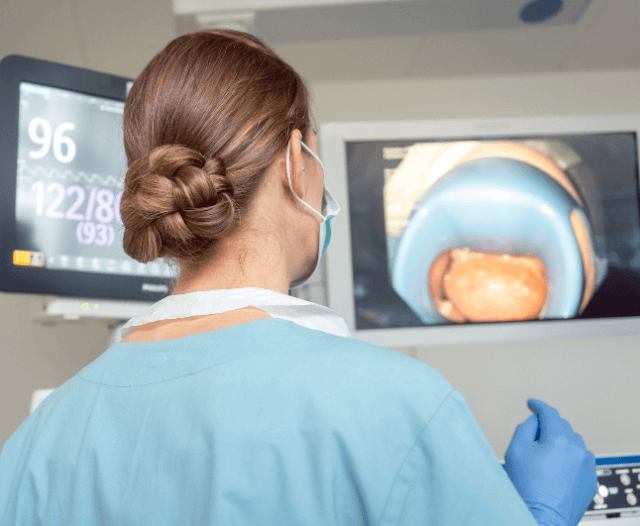
The camera transmits real-time images to a monitor, allowing the healthcare team to examine the lining of the digestive organs.


Visual Examination:

The gastroenterologist will carefully navigate the endoscope through the esophagus, stomach, and the initial part of the small intestine.

They will inspect the mucosal lining for abnormalities such as inflammation, ulcers, tumors, or other structural issues.


Biopsy (if necessary):

During the procedure, the gastroenterologist may take tissue samples (biopsies) for further examination.

This is a routine and relatively painless process. Biopsies can provide valuable information about the presence of infections, inflammation, or abnormalities.


Air Insufflation:

To enhance visibility, air may be gently introduced through the endoscope into the digestive tract. This can cause a temporary feeling of fullness or bloating.


Completion of Procedure:

Once the examination is complete, the endoscope will be carefully removed. You will be monitored as you recover from the sedation.

After the Procedure


Recovery Area:

You will be taken to a recovery area where you will be monitored as the sedation wears off. It is common to experience a mild sore throat or bloating due to the air insufflation.


Post-Procedure Discussion:

The gastroenterologist will meet with you to discuss their initial findings and share any immediate insights. If biopsies were taken, the results may take a few days and will be communicated to you in a follow-up appointment.


Rest and Recovery:

It’s advisable to rest for the remainder of the day. You should refrain from driving or making important decisions until the effects of the sedation wear off completely.


Dietary Guidelines:

You may be advised to start with a light diet and gradually resume your regular eating habits. Avoiding hot or spicy foods, as well as heavy or greasy meals, can help ease any residual discomfort.


Follow-Up Appointments:

Your healthcare provider will schedule a follow-up appointment to discuss the complete results of the gastroscopy, including any biopsy findings, and to determine the next steps in your care.

gastroscopy_aftercare


Potential Risks and Complications


While gastroscopy is generally considered safe, it is important to be aware of potential risks and complications associated with the procedure:

  • Sedation Risks: The use of sedative medications during gastroscopy carries a risk of complications, such as respiratory depression, cardiovascular events, or allergic reactions. However, these risks are minimal and are carefully monitored by the healthcare team.


  • Bleeding and Perforation: In rare cases, gastroscopy may cause bleeding or perforation of the gastrointestinal tract, particularly if therapeutic interventions are performed. However, the risk of these complications is low, especially when the procedure is performed by experienced gastroenterologists.


  • Discomfort: Some patients may experience mild discomfort or a gagging sensation during the insertion of the endoscope. However, sedation is typically administered to minimize discomfort and ensure patient comfort throughout the procedure.


After the Procedure


After the gastroscopy, you will be taken to a recovery area where you will be monitored as the effects of the sedation wear off.

You may experience some temporary side effects, such as a sore throat, bloating, or mild discomfort.

The healthcare team will provide instructions for post-procedure care, including dietary guidelines, activity restrictions, and when to follow up with your healthcare provider.


Conclusion


Navigating the unknowns of a gastroscopy procedure becomes more manageable with a comprehensive understanding of each step.

From the pre-procedure preparations to the recovery process, being informed empowers you to actively participate in your healthcare journey.

If you have any concerns or questions about the procedure, do not hesitate to discuss them with your healthcare provider.

Remember, the goal of a gastroscopy is to provide valuable insights into your digestive health, helping guide further diagnosis and treatment decisions.


gastroscopy_infographic


Contact Dr. Deetlefs


Dr. Deetlefs has earned the reputation of a trusted health expert providing consultative, diagnostic, and therapeutic endoscopic and related services to patients in Cape Town and beyond. 

Dr. Deetlefs can be contacted to confirm the estimated breakdown should you be interested in having the procedure done.

DISCLAIMER: PLEASE READ CAREFULLY

The information on this website is to provide general guidance. In no way does any of the information provided reflect definitive medical advice and self-diagnoses should not be made based on information obtained online. It is important to consult a Gastroenterologist or medical doctor regarding ANY and ALL symptoms or signs including, but not limited to: abdominal pain, haemorrhoids or anal / rectal bleeding as it may a sign of a serious illness or condition. A thorough consultation and examination should ALWAYS be performed for an accurate diagnosis and treatment plan. Be sure to call a physician or call our office today and schedule a consultation.

GIDoc Cape Town

Patient-focused GI treatments and procedures in Cape Town.

Monday-Friday 8AM-4PM.

Connect with Us

© Dr. Eduan Deetlefs, Registered Gastroenterologist, GI Doc Cape Town

Our website information is not intended or implied to be a substitute for professional medical advice, diagnosis or treatment. Please consult a doctor about your specific condition. Only a trained physician can determine an accurate diagnosis and proper treatment.

Polypectomy: Navigating the Landscape of Precancerous Lesion Removal

Polypectomy: Navigating the Landscape of Precancerous Lesion Removal


Polyps, abnormal tissue growths that can develop in various parts of the body, are commonly found in the colon.

When detected during a colonoscopy, these polyps may be removed through a procedure known as polypectomy.

This comprehensive guide aims to delve into the intricacies of polypectomy, exploring its purpose, the types of polyps encountered, the procedure itself, potential risks, and the significance of this intervention in preventing colorectal cancer.


Introduction


Polyps, although often benign, have the potential to transform into cancerous growths, particularly in the colon.

The importance of polypectomy lies in its role as a preventive measure, removing these precancerous lesions before they can progress.

This guide navigates through the world of polypectomy, shedding light on its significance in colorectal health.


Understanding Polyps and Their Types


Before delving into the specifics of polypectomy, it’s essential to understand the nature of polyps.

There are various types, including adenomatous polyps, hyperplastic polyps, and serrated polyps.

While most polyps are noncancerous, certain types have a higher likelihood of developing into colorectal cancer, underscoring the importance of early detection and removal.


Types of Polyps:

Polyps are abnormal tissue growths that can develop in various parts of the body, and they are commonly found in the colon.

The types of polyps can vary based on their characteristics and potential for malignancy. Here are some of the main types of polyps:


  1. Adenomatous Polyps:
  • Description:
    Adenomatous polyps are the most common type of polyps found in the colon.
  • Characteristics:
    They can be further classified into three subtypes – tubular, tubulovillous, and villous
    adenomas – based on their structure. Villous adenomas have a higher risk of developing into cancer.
  • Potential for Malignancy:
    Adenomatous polyps are considered precancerous, and their removal is crucial in preventing the development of colorectal cancer.


  1. Hyperplastic Polyps:
  • Description:
    Hyperplastic polyps are generally noncancerous and are more common in the rectum.
  • Characteristics:
    They are typically small and have a smoother appearance compared to adenomatous polyps.
  • Potential for Malignancy:
    While hyperplastic polyps themselves are not considered precancerous, some subtypes, especially those in the serrated category, may have an increased risk of developing into cancer.


  1. Serrated Polyps:
  • Description:
    Serrated polyps have a saw-toothed appearance under the microscope.
  • Characteristics: This category includes various types, such as traditional serrated adenomas, sessile serrated adenomas, and hyperplastic polyps with serrated features.
  • Potential for Malignancy: Some serrated polyps, particularly sessile serrated adenomas, are associated with an increased risk of developing into cancer.


  1. Inflammatory Polyps:
  • Description:
    Inflammatory polyps are typically associated with conditions such as inflammatory bowel disease (IBD).
  • Characteristics:
    They result from the body’s response to inflammation in the colon.
  • Potential for Malignancy:
    Inflammatory polyps themselves are not considered precancerous, but the underlying inflammation may increase the risk of developing colorectal cancer.


  1. Juvenile Polyps:
  • Description:
    Juvenile polyps are rare and are usually found in children.
  • Characteristics:
    They are typically small, and their appearance is distinctive under the microscope.
  • Potential for Malignancy:
    Juvenile polyps are generally noncancerous, but they can cause rectal bleeding and other symptoms.


  1. Peutz-Jeghers Polyps:
  • Description:
    Peutz-Jeghers polyps are associated with a rare genetic condition called Peutz-Jeghers syndrome.
  • Characteristics:
    They are often found in the stomach and small intestine.
  • Potential for Malignancy:
    While the polyps themselves are benign, individuals with Peutz-Jeghers syndrome have an increased risk of various cancers.


It’s important to note that the potential for malignancy varies among different types of polyps.

Regular screenings, such as colonoscopies, are crucial for detecting and removing precancerous polyps, thus preventing the development of colorectal cancer.

Individuals should discuss their specific situation with their healthcare provider to determine the most appropriate screening and surveillance plan based on their risk factors and medical history.


polypectomy_procedure_early_detection

The Importance of Early Detection


Early detection of polyps is paramount in preventing the progression to colorectal cancer.

Regular screenings, such as colonoscopies, play a crucial role in identifying and characterizing polyps, enabling timely intervention through polypectomy.


Polypectomy Procedure: Step by Step

Polypectomy is a vital medical procedure performed during a colonoscopy to remove polyps found in the colon.

These polyps, although often benign, have the potential to develop into colorectal cancer.

The step-by-step guide below provides a comprehensive overview of the polypectomy procedure, emphasizing its significance in preventing the progression of precancerous lesions.


Preparation:

Before the polypectomy procedure, the individual undergoes thorough preparation, typically involving a clear liquid diet and bowel cleansing to ensure optimal visualization of the colon during the colonoscopy.


Anesthesia and Sedation:

Once the patient is prepared, the healthcare provider administers anesthesia or sedation to ensure the patient is comfortable and relaxed during the procedure. In some cases, conscious sedation is used, allowing the patient to remain awake but in a relaxed state.


Insertion of Colonoscope:

The gastroenterologist or endoscopist then inserts a colonoscope into the rectum. A colonoscope is a flexible tube with a light and camera at its tip, allowing the healthcare provider to visualize the inside of the colon on a monitor.


Identification of Polyps:

As the colonoscope is navigated through the colon, the healthcare provider carefully examines the lining for the presence of polyps. Polyps may vary in size and shape, and their characteristics influence the choice of the polypectomy technique.


Snare Polypectomy:

For smaller polyps, a common technique is the snare polypectomy. The gastroenterologist uses a thin wire loop (snare) at the end of the colonoscope to encircle the polyp. Once the polyp is captured, the snare is tightened, cutting the polyp from the colon wall.


Endoscopic Mucosal Resection (EMR):

Larger polyps may require a more intricate technique known as Endoscopic Mucosal Resection (EMR). This technique involves injecting a saline solution beneath the polyp to lift it away from the colon wall. The elevated polyp is then snared and removed.


Electrocautery:

In some cases, particularly if bleeding is a concern, electrocautery may be used during the polypectomy. This involves applying an electrical current to the snare, coagulating blood vessels and minimizing the risk of bleeding.


Removal and Retrieval:

Once the polyp is successfully snared or resected, it is carefully removed from the colon. Larger polyps may be cut into smaller pieces for easier retrieval.


Biopsy (if needed):

In certain situations, the healthcare provider may perform a biopsy of the surrounding tissue to ensure complete removal of the polyp and to assess for any potential malignancy.


Post-Polypectomy Inspection:

After the polypectomy, the gastroenterologist carefully inspects the treated area to ensure there is no bleeding or other complications. Any potential bleeding can be addressed immediately during the procedure.


Recovery and Post-Procedure Care:

Following the polypectomy, the patient is monitored in a recovery area until the effects of sedation wear off. Instructions are provided regarding diet, activity, and any signs or symptoms that should prompt a follow-up.


types_of_polyps


Types of Polypectomy Techniques


Various techniques are utilized in polypectomy, each tailored to the characteristics of the polyp.

Snare polypectomy involves using a wire loop to snare and remove the polyp, while EMR is employed for larger lesions. This section explores the nuances of these techniques and their application.


Beyond Polypectomy: Colorectal Health and Cancer Prevention


Polypectomy is a pivotal intervention in colorectal health, but it is just one aspect of a broader strategy for preventing colorectal cancer.

Lifestyle factors, regular screenings, and awareness play integral roles in maintaining optimal colorectal health.


polypectomy_procedure_infographic


Contact Dr. Deetlefs


In conclusion, polypectomy emerges as a powerful tool in the prevention of colorectal cancer.

By understanding the procedure, its importance, and the collaborative role of individuals and healthcare providers, we empower ourselves in the journey toward optimal colorectal health.

Dr Eduan prides himself on his ability to help his patients to the best of his ability by embracing good listening skills, effective communication, compassion and knowledge and skill honed during years of private gastroenterology practice.

If you would like to book an appointment with a gastrointestinal (GI) specialist or would simply like more information on a particular GI topic, don’t hesitate to use our online booking form or call Dr. Deetlefs at 021 551 867. 

DISCLAIMER: PLEASE READ CAREFULLY

The information on this website is to provide general guidance. In no way does any of the information provided reflect definitive medical advice and self-diagnoses should not be made based on information obtained online. It is important to consult a Gastroenterologist or medical doctor regarding ANY and ALL symptoms or signs including, but not limited to: abdominal pain, haemorrhoids or anal / rectal bleeding as it may a sign of a serious illness or condition. A thorough consultation and examination should ALWAYS be performed for an accurate diagnosis and treatment plan. Be sure to call a physician or call our office today and schedule a consultation.

GIDoc Cape Town

Patient-focused GI treatments and procedures in Cape Town.

Monday-Friday 8AM-4PM.

Connect with Us

© Dr. Eduan Deetlefs, Registered Gastroenterologist, GI Doc Cape Town

Our website information is not intended or implied to be a substitute for professional medical advice, diagnosis or treatment. Please consult a doctor about your specific condition. Only a trained physician can determine an accurate diagnosis and proper treatment.

Ironing Out the Details: A Comprehensive Guide to Iron Deficiency Anemia

Ironing Out the Details: A Comprehensive Guide to Iron Deficiency Anemia

Overview


Iron deficiency anemia is one of the most common nutritional deficiencies worldwide, affecting millions of people.

Although it’s a condition often associated with fatigue, it presents a range of symptoms and can significantly affect an individual’s quality of life.

This blog post aims to delve into the causes, symptoms, and treatments of iron deficiency anemia, helping you understand this prevalent condition better.


What is Iron Deficiency Anemia?


Iron deficiency anemia is a condition characterized by a lack of healthy red blood cells due to insufficient iron in the body.

Iron is crucial for producing hemoglobin, a protein in red blood cells that enables them to carry oxygen around the body.

When your body lacks iron, it cannot produce enough hemoglobin, leading to decreased oxygen supply to your tissues, causing symptoms like fatigue, weakness, and difficulty concentrating.


Causes of Iron Deficiency Anemia


Several factors can lead to iron deficiency anemia:

  • Inadequate dietary intake: Our primary source of iron is our diet. Foods rich in iron include meat, eggs, green leafy vegetables, and iron-fortified cereals. A diet lacking these can result in an iron deficiency. 
  • Absorption issues: Conditions such as celiac disease or surgical procedures that remove parts of the stomach or small intestine can hinder the body’s ability to absorb iron efficiently. 
  • Pregnancy: Pregnant women require significantly more iron to support their growing babies and the increase in their blood volume. Without adequate iron supplementation, this could lead to deficiency. 
  • Blood loss: Conditions that cause blood loss, such as heavy menstrual periods or chronic diseases like peptic ulcers and colorectal cancer, can deplete the body’s iron stores.


iron_deficiency_anemia_symptoms


Symptoms of Iron Deficiency Anemia


Iron deficiency anemia manifests in various ways, including:

  • Fatigue and weakness: This is often the first sign of anemia, as your tissues aren’t getting enough oxygen, causing you to feel constantly tired or weak. 
  • Pale skin: Hemoglobin gives skin its rosy color, so low levels can cause your skin to become lighter. 
  • Shortness of breath: This symptom occurs because your body attempts to increase oxygen delivery to your tissues. 
  • Headaches and dizziness: Insufficient oxygen reaching the brain can cause these symptoms. 
  • Unusual cravings: Some people with iron deficiency anemia develop cravings for non-food items like ice or dirt, a condition known as pica. 
  • Restless legs syndrome: Some people with iron deficiency experience an overwhelming urge to move their legs, particularly at night.

Diagnosing Iron Deficiency Anemia


Iron deficiency anemia is diagnosed through a combination of medical history, physical examination, and laboratory tests.

Here are the typical steps in diagnosing this condition:

Medical History and Physical Examination:
Your doctor will start by asking about your symptoms, diet, family history, and any medicines you’re taking. They will also conduct a physical examination to look for signs of iron deficiency anemia such as pale skin, a fast or irregular heartbeat, or a swollen tongue.

Blood Tests:
The main diagnostic tool for iron deficiency anemia is blood testing.

Here are some of the most common tests used:

Complete Blood Count (CBC):
This test measures different components of your blood, including hemoglobin and hematocrit levels, as well as the size and shape of your red blood cells. In iron deficiency anemia, the hemoglobin level is low, and the red blood cells are usually smaller and paler than normal.

Serum Iron and Ferritin Tests: These tests measure the amount of iron in your blood and the amount of stored iron in your body, respectively. Both are typically low in iron deficiency anemia.

Transferrin Level and Total Iron-Binding Capacity (TIBC): Transferrin is a protein that carries iron in your blood, and TIBC measures how well your body can bind iron. In iron deficiency anemia, transferrin levels are high and TIBC is increased, as your body tries to maximize iron uptake.

Further Testing: If the cause of your iron deficiency anemia isn’t clear, your doctor might recommend further testing. This could include endoscopy to check for sources of internal bleeding, colonoscopy to look for bleeding in the colon, or tests to look for celiac disease, which can interfere with the absorption of iron from your diet.

Remember that iron deficiency anemia is a treatable condition, but it’s important to identify and address the underlying cause.

If you suspect you have iron deficiency anemia, it’s important to consult a healthcare provider for a proper diagnosis and treatment plan.

anemia_diagnosis


Dealing With Iron Deficiency on A Daily Basis

Living with iron deficiency anemia can be challenging due to the fatigue and other symptoms associated with the condition.

Here are some strategies to manage and cope with iron deficiency anemia on a daily basis:

Follow your treatment plan: Adhere to the treatment plan set out by your doctor. This might include taking iron supplements, eating an iron-rich diet, or treating underlying medical conditions. Make sure to keep all medical appointments and contact your healthcare provider if you experience any problems with your treatment.

Eat a balanced, iron-rich diet: Include iron-rich foods in your daily diet. Lean meats, poultry, fish, iron-fortified cereals and breads, dark green leafy vegetables, beans, and lentils are all excellent sources of dietary iron. Foods high in vitamin C, such as citrus fruits, strawberries, and bell peppers, can help enhance iron absorption.

Limit intake of foods that hinder iron absorption: Certain foods and beverages, such as coffee, tea, dairy products, whole grains, and some legumes, can inhibit iron absorption if consumed at the same time as iron-rich foods or supplements.

Exercise regularly: Although it may be challenging when you’re feeling fatigued, regular moderate exercise can help stimulate your appetite, promote good sleep, and boost your energy levels. Start small and gradually increase your activity as your energy levels improve.

Stay hydrated: Drinking plenty of fluids can help combat symptoms of fatigue and help with the constipation that can sometimes occur with iron supplements.

Manage stress: Chronic stress can exacerbate symptoms of anemia. Techniques such as mindfulness, deep breathing, yoga, and other relaxation exercises can help manage stress levels.

Monitor your menstrual cycle: If you are a woman with heavy menstrual bleeding, discuss this with your doctor, as it can contribute to iron deficiency anemia. There may be treatment options to manage heavy periods.

Stay connected with your healthcare team: Regular follow-ups with your healthcare provider are essential to monitor your condition and adjust treatment as needed.

Remember that recovery from iron deficiency anemia often takes time and requires consistent effort. Over time, with consistent treatment and healthy lifestyle habits, you should see improvement in your symptoms.


iron_deficiency_anemia_guide_infogrpahic


Conclusion


Despite the challenges, it is reassuring to know that iron deficiency anemia is generally treatable, and most individuals experience a significant improvement in their symptoms with appropriate management.

By adhering to recommended treatments, maintaining an iron-rich diet, and staying connected with their healthcare providers, individuals with this condition can lead a healthy, fulfilling life.

For any additional information regarding iron deficiency anemia, it’s best to find a trusted and experienced Gastroenterologist who can answer your questions clearly, recommend appropriate solutions, and perform tests with precision and expertise.

Dr. Deetlefs has earned the reputation of a trusted health expert providing consultative, diagnostic, and therapeutic endoscopic and related services to patients in Cape Town and beyond.

Dr Eduan prides himself on his ability to help his patients to the best of his ability by embracing good listening skills, effective communication, compassion and knowledge and skill honed during years of private gastroenterology practice.

If you would like to book an appointment with a gastrointestinal (GI) specialist or would simply like more information on a particular GI topic, don’t hesitate to use our online booking form
or call Dr. Deetlefs at 021 551 867.

DISCLAIMER: PLEASE READ CAREFULLY

The information on this website is to provide general guidance. In no way does any of the information provided reflect definitive medical advice and self-diagnoses should not be made based on information obtained online. It is important to consult a Gastroenterologist or medical doctor regarding ANY and ALL symptoms or signs including, but not limited to: abdominal pain, haemorrhoids or anal / rectal bleeding as it may a sign of a serious illness or condition. A thorough consultation and examination should ALWAYS be performed for an accurate diagnosis and treatment plan. Be sure to call a physician or call our office today and schedule a consultation.

GIDoc Cape Town

Patient-focused GI treatments and procedures in Cape Town.

Monday-Friday 8AM-4PM.

Connect with Us

© Dr. Eduan Deetlefs, Registered Gastroenterologist, GI Doc Cape Town

Our website information is not intended or implied to be a substitute for professional medical advice, diagnosis or treatment. Please consult a doctor about your specific condition. Only a trained physician can determine an accurate diagnosis and proper treatment.

Unraveling the Mystery of Crohn’s Disease: Causes, Symptoms, and Insights

Unraveling the Mystery of Crohn’s Disease: Causes, Symptoms, and Insights

Gain a better understanding of Crohn’s disease, a chronic inflammatory bowel condition that can affect various parts of the digestive tract.

Delve into the potential causes, common symptoms, and the diagnostic process to understand this complex autoimmune disorder better.

What is Crohn’s Disease?

Crohn’s disease is a chronic inflammatory bowel disease (IBD) that affects the lining of the digestive tract, also known as the gastrointestinal (GI) tract.

It can involve any part of the GI tract from the mouth to the anus, but it most commonly affects the end of the small intestine (ileum) and the beginning of the large intestine (colon).

The inflammation caused by Crohn’s disease can penetrate deep into the layers of the affected bowel tissue, leading to pain, discomfort, and various digestive symptoms.

The inflammation can also cause complications such as strictures (narrowing of the intestine), fistulas (abnormal connections between different parts of the intestine or between the intestine and other organs), and abscesses (pockets of infection).

Causes and Risk Factors

The exact cause of Crohn’s disease is still not completely understood, but it is believed to result from a complex interplay of genetic, environmental, and immune system factors.

Here are some of the known causes and risk factors associated with Crohn’s disease:

Genetics:
There is a clear genetic component to Crohn’s disease, as individuals with a family history of the condition are more likely to develop it. Several genes have been identified that may increase the risk of developing Crohn’s disease, but not everyone who carries these genes will develop the condition.
 

Immune system:
Crohn’s disease is considered an autoimmune disorder, in which the immune system mistakenly attacks healthy cells in the gastrointestinal tract. This immune response leads to chronic inflammation and tissue damage. The exact reason for this abnormal immune response is not fully understood, but it is believed to involve a combination of genetic and environmental factors.

Environmental factors:
Although the exact environmental triggers of Crohn’s disease are not known, several factors have been associated with an increased risk of developing the condition. These factors include smoking and having a diet high in fat and low in fiber. It has also been suggested that infections, particularly those involving the gastrointestinal tract, may play a role in triggering Crohn’s disease in susceptible individuals.

Age:
Crohn’s disease can occur at any age, but it is most commonly diagnosed in young adults between the ages of 15 and 35. However, it is important to note that the condition can develop later in life as well.

Smoking:
Smoking is a significant risk factor for the development of Crohn’s disease and is also associated with more severe disease and a higher risk of complications. Quitting smoking can help reduce the risk of developing the condition and improve the overall health of those who already have it.

crohns_symptoms

Common Symptoms of Crohn’s

The symptoms of Crohn’s disease can vary widely depending on the severity of inflammation and the specific parts of the digestive tract affected. Some common symptoms associated with Crohn’s disease include:

Abdominal Pain and Cramping:
This is often experienced in the lower right part of the abdomen but can occur anywhere along the digestive tract. The pain may be mild to severe and can come and go, often worsening after meals.

Diarrhea:
Frequent loose and watery stools are a common symptom of Crohn’s disease. In some cases, diarrhea may be severe and accompanied by blood or mucus.

Fatigue:
People with Crohn’s disease often experience a general feeling of tiredness and low energy levels due to inflammation, malabsorption of nutrients, and the impact of other symptoms on daily life.

Weight loss:
Unintentional weight loss can result from a combination of reduced appetite, malabsorption of nutrients, and increased calorie expenditure due to the ongoing inflammation in the digestive tract.

Fever:
A low-grade fever can sometimes accompany active inflammation in Crohn’s disease, particularly during flare-ups.

Mouth sores:
Painful sores, or ulcers, can develop in the mouth as a result of inflammation in the digestive tract.

Perianal disease:
This includes symptoms like pain, swelling, or drainage around the anus, which can result from the formation of abscesses, fissures, or fistulas.

Nutritional deficiencies:
Crohn’s disease can cause malabsorption of nutrients, leading to deficiencies in essential vitamins and minerals such as iron, vitamin B12, and vitamin D.

Joint pain:
Arthritis or joint pain can occur in some individuals with Crohn’s disease, affecting the knees, ankles, wrists, or other joints.

Skin issues:
Some people with Crohn’s disease may develop skin problems, such as erythema nodosum (painful red nodules under the skin) or pyoderma gangrenosum (painful ulcers that develop rapidly).


Eye inflammation:
Inflammation of the eyes, such as uveitis or episcleritis, can also be a symptom of Crohn’s disease.

crohns_diagnosis_and_testing


Diagnosis and Testing

Diagnosing Crohn’s disease can be challenging due to its varied symptoms and the fact that it can mimic other gastrointestinal conditions.

A combination of medical history, physical examination, and various tests and procedures is often required to accurately diagnose Crohn’s disease.

Here are some of the common steps and tests involved in diagnosing the condition:

Medical History and Physical Examination:
The physician will begin by taking a detailed medical history, including information about symptoms, family history of Crohn’s disease or other gastrointestinal disorders, and any medications the patient is taking. A physical examination will follow, focusing on signs of inflammation, malnutrition, or complications related to Crohn’s disease.

Blood Tests:
Blood tests can help identify signs of inflammation, anemia, and possible nutritional deficiencies. Common blood tests include complete blood count (CBC), C-reactive protein (CRP), and erythrocyte sedimentation rate (ESR).

Stool Tests:
Stool samples may be analyzed to rule out other causes of gastrointestinal symptoms, such as infections or parasites. The presence of fecal calprotectin, a protein that indicates inflammation in the intestines, can also be detected in stool samples and may be a marker for Crohn’s disease.

Imaging Studies:
Various imaging tests can help visualize the gastrointestinal tract and identify areas of inflammation, strictures, or other abnormalities. These tests may include X-rays, computed tomography (CT) scans, magnetic resonance imaging (MRI), or ultrasound.
 

Endoscopy:
Endoscopic procedures involve inserting a flexible tube with a camera at its tip into the gastrointestinal tract to directly visualize the lining of the digestive tract and take biopsies (tissue samples) for further analysis. Two common endoscopic procedures used in diagnosing Crohn’s disease are:

Colonoscopy:
This procedure allows the physician to examine the entire colon and the end of the small intestine (ileum). Biopsies can be taken during a colonoscopy to help confirm the diagnosis.

upper_endoscopy_procedure

Upper endoscopy:
This procedure is used to examine the esophagus, stomach, and the first part of the small intestine (duodenum). It may be performed if symptoms suggest Crohn’s disease is affecting the upper gastrointestinal tract.

Capsule endoscopy:
In some cases, a capsule endoscopy may be recommended to visualize the small intestine, which can be difficult to examine using conventional endoscopy. The patient swallows a small capsule containing a camera, which takes pictures of the digestive tract as it passes through, transmitting the images to a recording device worn by the patient.

mystery_of_crohns_infographic

Conclusion

Dr. Deetlefs has earned the reputation of a trusted health expert providing consultative, diagnostic, and therapeutic endoscopic and related services to patients in Cape Town and beyond.

Dr Eduan prides himself on his ability to help his patients to the best of his ability by embracing good listening skills, effective communication, compassion and knowledge and skill honed during years of private gastroenterology practice.

If you would like to book an appointment with a gastrointestinal (GI) specialist or would simply like more information on a particular GI topic, don’t hesitate to use our online booking form
or call Dr. Deetlefs at 021 551 867. 

DISCLAIMER: PLEASE READ CAREFULLY

The information on this website is to provide general guidance. In no way does any of the information provided reflect definitive medical advice and self-diagnoses should not be made based on information obtained online. It is important to consult a Gastroenterologist or medical doctor regarding ANY and ALL symptoms or signs including, but not limited to: abdominal pain, haemorrhoids or anal / rectal bleeding as it may a sign of a serious illness or condition. A thorough consultation and examination should ALWAYS be performed for an accurate diagnosis and treatment plan. Be sure to call a physician or call our office today and schedule a consultation.

GIDoc Cape Town

Patient-focused GI treatments and procedures in Cape Town.

Monday-Friday 8AM-4PM.

Connect with Us

© Dr. Eduan Deetlefs, Registered Gastroenterologist, GI Doc Cape Town

Our website information is not intended or implied to be a substitute for professional medical advice, diagnosis or treatment. Please consult a doctor about your specific condition. Only a trained physician can determine an accurate diagnosis and proper treatment.

Understanding Ulcerative Colitis

Understanding Ulcerative Colitis

Overview of Ulcerative Colitis


Ulcerative colitis is a chronic inflammatory bowel disease that primarily affects the colon and rectum. The condition causes inflammation and sores (ulcers) to develop on the inner lining of the large intestine, leading to symptoms such as diarrhea, abdominal pain, and rectal bleeding.

The exact cause of ulcerative colitis is not known, but it is thought to be an autoimmune disorder in which the body’s immune system mistakenly attacks the lining of the colon and rectum.

Other factors that may contribute to the development of ulcerative colitis include genetics, environmental factors, and an imbalance of gut bacteria.

Symptoms of ulcerative colitis can vary in severity and may include diarrhea, bloody stools, abdominal pain and cramping, fatigue, weight loss, and fever.

Flares of the disease can be unpredictable and may be triggered by factors such as stress, infections, or changes in diet.

Treatment for ulcerative colitis typically involves medications to reduce inflammation and manage symptoms, such as corticosteroids, immunosuppressants, and biologics.

In this blog, we will delve into the topic of ulcerative colitis, a chronic inflammatory bowel disease that affects the colon and rectum.


Symptoms


The symptoms of ulcerative colitis can vary from person to person, and they may range from mild to severe.

Some of the most common symptoms of UC include:

  • Diarrhea, often with blood or pus
  • Abdominal pain and cramping
  • Urgency to have a bowel movement
  • Inability to have a bowel movement despite urgency
  • Rectal pain and bleeding
  • Fatigue
  • Loss of appetite and weight loss
  • Anemia

ulcerative_colitis_diagnosis


Causes


The exact cause of ulcerative colitis is unknown, but it is thought to be a result of a combination of genetic, environmental, and immunological factors.

People with a family history of UC are at a higher risk of developing the disease.

Other factors that may contribute to the development of UC include a compromised immune system, bacterial or viral infections, and stress.


Diagnosis

The diagnosis of ulcerative colitis involves a combination of medical history, physical examination, and diagnostic tests.

A healthcare provider will ask about the patient’s symptoms, medical history, and family history of IBD.
A physical exam may also be conducted to check for signs of inflammation in the abdomen.

Diagnostic tests that may be used to diagnose UC include: 

Blood tests to check for signs of inflammation or infection

Blood tests can be useful in the diagnosis and management of ulcerative colitis (UC).
While there is no one specific blood test that can definitively diagnose UC, blood tests can help rule out other conditions that may have similar symptoms and help monitor disease activity and response to treatment.

Some of the tests that may be ordered for UC include:

  • Complete blood count (CBC):
    This test measures the levels of different types of blood cells, including red blood cells, white blood cells, and platelets. In UC, the CBC can show signs of anemia (low red blood cell count) or inflammation (elevated white blood cell count).
  • Stool tests to check for blood, infections, or parasites
  • CT scan or MRI to evaluate the extent of inflammation and complications
  • Colonoscopy to visualize the colon and take tissue samples for biopsy

ulcerative_colitis_treatment


Colonoscopy Explained


Colonoscopy is a medical procedure that allows healthcare providers to visualize the inside of the colon and rectum using a flexible, lighted tube called a colonoscope. 

The procedure is used to screen for colon cancer, diagnose and treat various gastrointestinal conditions, and monitor the effectiveness of treatment.

During a colonoscopy, the patient lies on their side while the colonoscope is inserted into the rectum and advanced through the colon.

The colonoscope has a tiny camera on the end, which sends images to a monitor that the healthcare provider uses to examine the colon lining for abnormalities such as polyps, inflammation, or cancerous growths.

Before a colonoscopy, patients must follow a special diet and take laxatives to cleanse the colon.

They may also receive sedation or anesthesia to help them relax and reduce discomfort during the procedure.

If any abnormalities are found during the colonoscopy, the healthcare provider may take a biopsy or remove the abnormal tissue for further testing.

The removed tissue is then sent to a lab for analysis to determine if it is cancerous or benign.

Although colonoscopy is generally considered a safe procedure, there are some risks, including bleeding, infection, and perforation of the colon.

It is important to discuss any concerns with the healthcare provider before the procedure.

In summary, colonoscopy is a procedure that allows healthcare providers to examine the colon and rectum for abnormalities using a colonoscope.

The procedure is used to screen for conditions and diagnose and treat gastrointestinal conditions, and monitor treatment effectiveness.

Patients must prepare for the procedure by following a special diet and taking laxatives to cleanse the colon.

If abnormalities are found, a biopsy or removal of tissue may be necessary for further testing.

While generally safe, there are some risks associated with the procedure.


ulcerative_colitis_colonoscopy


Treatment


The treatment of ulcerative colitis may involve medications and lifestyle changes.
The goals of treatment are to reduce inflammation, manage symptoms, and prevent complications.

Medications that may be used to treat UC include: 

  • Aminosalicylates (5-ASAs) to reduce inflammation
  • Corticosteroids to reduce inflammation
  • Immunosuppressants to suppress the immune system
  • Biologic therapies to target specific proteins involved in inflammation
  • Lifestyle changes
  • Eating a healthy and balanced diet
  • Staying hydrated
  • Getting regular exercise
  • Reducing stress
  • Quitting smoking

In some cases, surgery may be necessary to remove the affected part of the colon or rectum.


Traveling with Ulcerative Colitis:


Traveling can be stressful for people with ulcerative colitis, as it can disrupt daily routines and access to medical care. 

It is important to stay calm during travelling, keep an emergency kit with you and stay hydrated.


Alternative Therapies for Ulcerative Colitis:

While conventional medical treatments are often effective for managing ulcerative colitis, some people may seek out alternative therapies, such as acupuncture or herbal remedies.


The Latest Research on Ulcerative Colitis:


Ulcerative colitis is a rapidly evolving field, with new research and treatments emerging all the time. 

In this blog post, you could highlight some of the latest research on ulcerative colitis, including advances in genetics, immunology, and microbiome research. 

ulcerative_infographic


Conclusion:


In conclusion, ulcerative colitis is a chronic inflammatory disease that affects the colon and rectum.

It can cause a range of uncomfortable symptoms.  Although there is no known cure for ulcerative colitis, there are a variety of treatment options available to manage the symptoms and improve quality of life for patients.

It is important for individuals with ulcerative colitis to work closely with their healthcare providers to develop a personalized treatment plan that addresses their specific symptoms and needs. This may involve a combination of medications, lifestyle changes, and dietary modifications.

Furthermore, it is essential to stay up to date with regular check-ups and monitoring to ensure that the disease is under control and to prevent the development of serious complications, such as colon cancer.

Living with ulcerative colitis can be challenging, but with proper care and management, it is possible to lead a healthy and fulfilling life.

For any additional information regarding a Colonoscopy procedure, it’s best to find a trusted and experienced Gastroenterologist who can answer your questions clearly, recommend appropriate solutions, and perform tests with precision and expertise.

Dr. Deetlefs has earned the reputation of a trusted health expert providing consultative, diagnostic, and therapeutic endoscopic and related services to patients in Cape Town and beyond.

Dr Eduan prides himself on his ability to help his patients to the best of his ability by embracing good listening skills, effective communication, compassion and knowledge and skill honed during years of private gastroenterology practice.


If you would like to book an appointment with a gastrointestinal (GI) specialist or would simply like more information on a particular GI topic, don’t hesitate to use our online booking form
or call Dr. Deetlefs at 021 551 867. 

DISCLAIMER: PLEASE READ CAREFULLY

The information on this website is to provide general guidance. In no way does any of the information provided reflect definitive medical advice and self-diagnoses should not be made based on information obtained online. It is important to consult a Gastroenterologist or medical doctor regarding ANY and ALL symptoms or signs including, but not limited to: abdominal pain, haemorrhoids or anal / rectal bleeding as it may a sign of a serious illness or condition. A thorough consultation and examination should ALWAYS be performed for an accurate diagnosis and treatment plan. Be sure to call a physician or call our office today and schedule a consultation.

GIDoc Cape Town

Patient-focused GI treatments and procedures in Cape Town.

Monday-Friday 8AM-4PM.

Connect with Us

© Dr. Eduan Deetlefs, Registered Gastroenterologist, GI Doc Cape Town

Our website information is not intended or implied to be a substitute for professional medical advice, diagnosis or treatment. Please consult a doctor about your specific condition. Only a trained physician can determine an accurate diagnosis and proper treatment.

Colonoscopy Explained

Colonoscopy Explained


What is a Colonoscopy?


A colonoscopy is a medical procedure used to examine the inside of the large intestine (colon) and rectum.

During the procedure, a long, flexible, and lighted instrument called a colonoscope is inserted through the rectum to visualize the entire colon.

The procedure is used to detect and diagnose conditions such as colon cancer, inflammatory bowel disease (IBD), diverticulosis, and to remove polyps or other growths.

Why is a Colonoscopy Important

A colonoscopy is important because it allows the doctor to visually examine the inside of the colon for any abnormalities such as polyps, tumors, or inflammation.

It can help diagnose various conditions such as colon cancer, inflammatory bowel disease (IBD), and diverticulosis.

It is also used as a screening tool to detect colon cancer in its early stages, when it is most treatable.

Additionally, if any abnormal growths are found during the procedure, they can be removed immediately, reducing the risk of future complications.


How to Prepare for a Colonoscopy

To prepare for a colonoscopy, you will need to follow specific instructions provided by your doctor to clean out your colon, which typically include:


  • Diet changes: Avoid solid foods for 1 – 2 days before the procedure and follow a clear liquid diet. 
  • Laxatives: Take prescribed or over-the counter laxatives to empty your bowels. 
  • Hydration: Drink plenty of liquids to stay hydrated 
  • Avoid certain medications: Stop taking certain medications such as blood thinners before procedure. 
  • Fast: Don’t eat or drink anything for 8 – 12 hours before the procedure. 

It’s important to follow the instructions provided by your doctor closely, as a clean colon is essential for a successful and safe colonoscopy.

Additionally, let your doctor know of any medications you are taking, and if you have any medical conditions or allergies.

colonoscopy_Prepare

What Happens During a Colonoscopy

During a colonoscopy, the patient is positioned on their side on an exam table and given sedation to relax and make the procedure more comfortable.

The doctor then inserts the colonoscope through the rectum and guides it through the colon.

The scope has a light and camera, allowing the doctor to see the inside of the colon and take images for later review.

If any polyps or other growths are found, the doctor can remove them during the procedure using instruments passed through the colonoscope.

The procedure typically lasts 30 minutes to an hour.

After the procedure, the patient should rest for a short time under observation, until the sedation wears off.

The patient may feel some bloating or mild cramping, but these symptoms typically resolve quickly.

The doctor will discuss the results of the colonoscopy with the patient, including any findings and the need for any further tests or treatments.


Risks and Potential Complications

A colonoscopy is a generally safe procedure, but like any medical procedure, there are some risks and potential complications.

These include:

  • Bleeding: There is a small risk of bleeding after the removal of a polyp or growth.
  • Perforation: In rare cases, the colonoscope may cause a tear in the colon, which can cause peritonitis (an infection of the abdominal cavity). 
  • Reaction to sedation: Some people may have an adverse reaction to the sedation used during the procedure. 
  • Discomfort: Some patients may experience discomfort or mild pain during or after the procedure. 
  • Missed lesions: While a colonoscopy is highly effective, it is not foolproof and there is a small risk that a lesion or growth may be missed.

It is important to discuss any concerns or questions about the risks and potential complications of a colonoscopy with your doctor before the procedure.

Your doctor can also explain what to expect during and after the procedure and answer any questions you may have.


colonoscopy_procedure


How to Care for Yourself After a Colonoscopy

After a colonoscopy, you should follow these steps to ensure a smooth recovery:

  • Rest: Take it easy for the rest of the day and avoid strenuous activities. 
  • Hydration: Drink plenty of clear liquids to stay hydrated. 
  • Resume normal activities: Gradually return to your normal diet and activities but avoid solid foods for a few hours after the procedure. 
  • Follow-up care: If you had any polyps or growths removed during the procedure, your doctor will let you know if any follow-up care is needed. 
  • Report any symptoms: If you experience any symptoms such as severe abdominal pain, fever, or heavy bleeding, contact your doctor immediately. 

It is important to follow your doctor’s instructions and listen to your body as you recover from a colonoscopy.

If you have any concerns or questions about your recovery, do not hesitate to reach out to your doctor for advice.


Alternatives to a Colonoscopy


There are several alternatives to a colonoscopy, including:

  • Fecal Occult Blood Test (FOBT): A simple blood test to detect blood in the stool, which can be a sign of colon cancer. 
  • Stool DNA test: A test that looks for abnormal DNA in the stool, which can indicate the presence of colon cancer or precancerous polyps. 
  • Flexible Sigmoidoscopy: Similar to a colonoscopy, but only examines the lower part of the colon. 
  • CT Colonography (Virtual Colonoscopy): A non-invasive test that uses X-rays and computer software to produce images of the colon. 
  • Double-Contrast Barium Enema (DCBE): An X-ray test that uses barium to highlight the inside of the colon.

It’s important to discuss your options with your doctor, who can help determine which test is best for you based on your individual needs and risk factors.

Keep in mind that while these tests can be used to detect colon cancer or precancerous growths, a colonoscopy remains the most effective way to examine the inside of the colon and remove any growths or polyps if necessary.

colonoscopy_diagnosis


When to Schedule Your Next Colonoscopy

The recommended interval for scheduling your next colonoscopy depends on several factors, including:

  • Age: For average-risk individuals, the American Cancer Society recommends a first colonoscopy at age 45 or earlier if there is a family history of colon cancer. 
  • Risk factors: If you have any risk factors for colon cancer, such as a family history, personal history of polyps or colon cancer, or inflammatory bowel disease, your doctor may recommend a more frequent screening schedule. 
  • Results of previous colonoscopies: If no polyps or growths were found during your previous colonoscopy, your doctor may recommend scheduling your next colonoscopy in 10 years.If polyps were found and removed, the recommended interval may be shorter, based on the size, number, and type of polyps.

It is important to discuss your individual screening schedule with your doctor, who can help determine the right time for your next colonoscopy based on your individual health history and risk factors.

colonocopy_explained_infographic


Conclusion

For any additional information regarding a Colonoscopy procedure, it’s best to find a trusted and experienced Gastroenterologist who can answer your questions clearly, recommend appropriate solutions, and perform tests with precision and expertise.

Dr. Deetlefs has earned the reputation of a trusted health expert providing consultative, diagnostic, and therapeutic endoscopic and related services to patients in Cape Town and beyond.

Dr Eduan prides himself on his ability to help his patients to the best of his ability by embracing good listening skills, effective communication, compassion and knowledge and skill honed during years of private gastroenterology practice.


If you would like to book an appointment with a gastrointestinal (GI) specialist or would simply like more information on a particular GI topic, don’t hesitate to use our online booking form
or call Dr. Deetlefs at 021 551 867. 

DISCLAIMER: PLEASE READ CAREFULLY

The information on this website is to provide general guidance. In no way does any of the information provided reflect definitive medical advice and self-diagnoses should not be made based on information obtained online. It is important to consult a Gastroenterologist or medical doctor regarding ANY and ALL symptoms or signs including, but not limited to: abdominal pain, haemorrhoids or anal / rectal bleeding as it may a sign of a serious illness or condition. A thorough consultation and examination should ALWAYS be performed for an accurate diagnosis and treatment plan. Be sure to call a physician or call our office today and schedule a consultation.

GIDoc Cape Town

Patient-focused GI treatments and procedures in Cape Town.

Monday-Friday 8AM-4PM.

Connect with Us

© Dr. Eduan Deetlefs, Registered Gastroenterologist, GI Doc Cape Town

Our website information is not intended or implied to be a substitute for professional medical advice, diagnosis or treatment. Please consult a doctor about your specific condition. Only a trained physician can determine an accurate diagnosis and proper treatment.