021 202 0626
PEG Feeding Tubes: When Are They Needed?

PEG Feeding Tubes: When Are They Needed?

When swallowing difficulties make it unsafe to eat, or illness prevents a person from meeting their nutritional needs, long-term nutrition may need to be delivered another way. A PEG feeding tube provides direct access to the stomach through the abdominal wall. It can support recovery, maintain hydration and body weight, and make it easier to give prescribed liquid nutrition or medicines. However, PEG placement is not appropriate for everyone, and it should follow a careful discussion of the patient’s diagnosis, expected benefit, overall health, values and goals of care.

A percutaneous endoscopic gastrostomy, usually shortened to PEG, is one of the advanced gastroenterology treatments and procedures offered at GI Doc Cape Town. This guide explains when it may be recommended, what happens during placement, how the tube is used and which symptoms require medical attention.

What Is a PEG Feeding Tube?

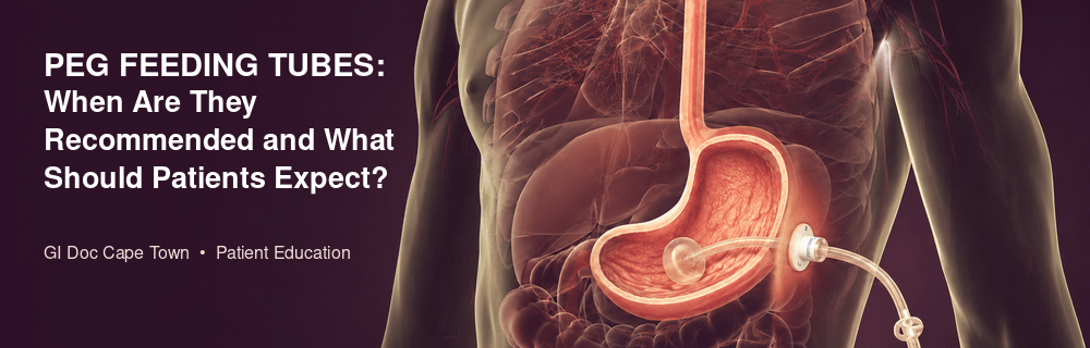
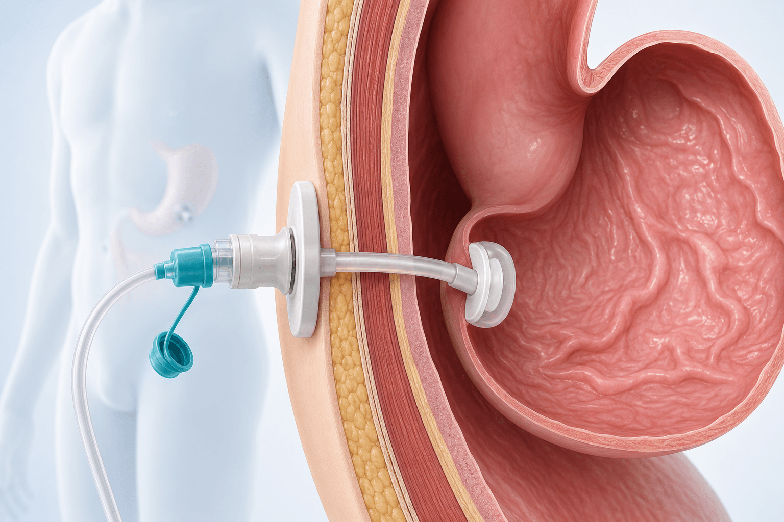
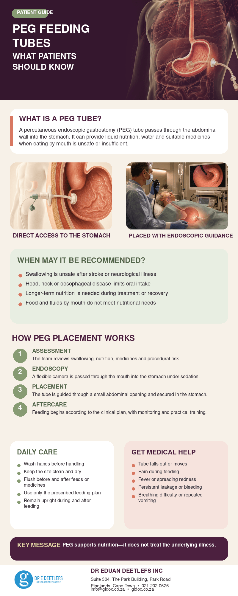
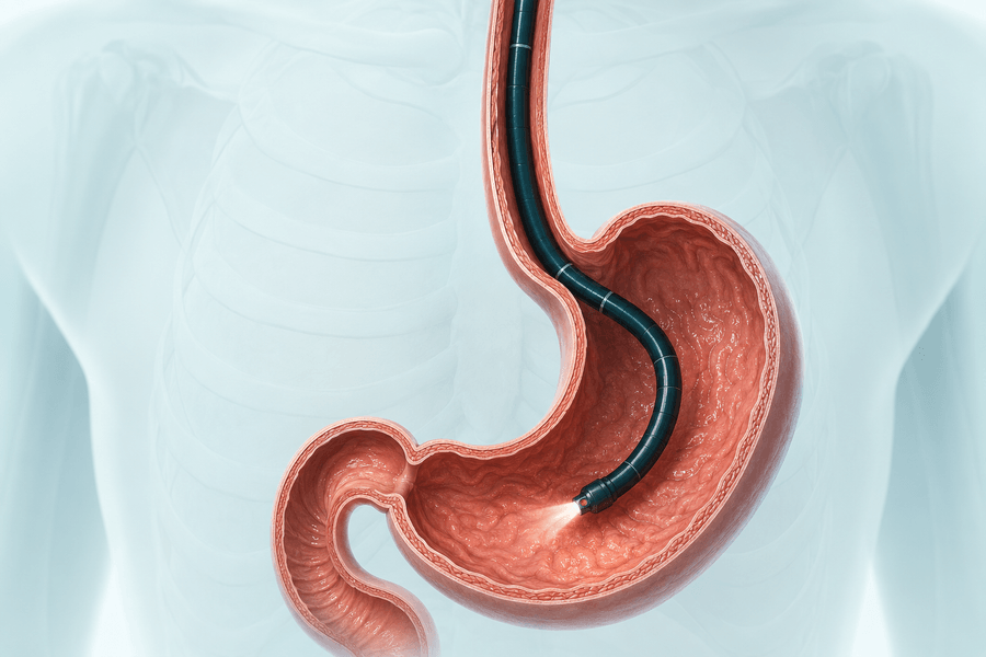
A PEG is a flexible tube placed through the skin of the abdomen and into the stomach using an endoscope. “Percutaneous” means through the skin, “endoscopic” refers to the flexible camera used to view the stomach, and “gastrostomy” describes the opening created into the stomach.

Once in position, the tube provides a route for specially formulated liquid nutrition, water and suitable medicines. It does not enter a vein. This is called enteral nutrition because nourishment still passes into the digestive tract, allowing the stomach and intestines to continue doing their work.

When May a PEG Be Recommended?

PEG placement may be discussed when a person cannot consume enough food and fluid safely, but the stomach and intestines can still absorb nutrition. The recommendation is based on the underlying condition, nutritional status, expected length of support and likelihood that the tube will provide meaningful benefit.

peg_tubes_procedure_3d

Neurological Conditions and Swallowing Problems

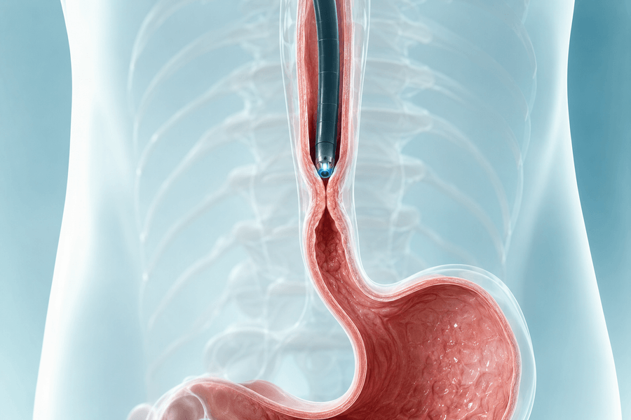
A stroke, traumatic brain injury, motor neurone disease, Parkinson’s disease, multiple sclerosis or another neurological condition may interfere with chewing and swallowing. Food or liquid can then enter the airway rather than the oesophagus, increasing the risk of aspiration and chest infection. A PEG may provide a safer route for nutrition while swallowing is assessed or rehabilitation continues.

Head, Neck or Oesophageal Disease

Tumours, surgery or radiotherapy involving the mouth, throat, head or neck may make eating painful, difficult or unsafe. Severe narrowing of the oesophagus can also obstruct the passage of food. In some cases, endoscopic treatment may improve the narrowing; in others, tube feeding is needed to protect nutrition before, during or after treatment. A gastroscopy may be part of the assessment of the oesophagus and stomach, depending on the clinical situation.

Severe Illness, Malnutrition or Prolonged Recovery

Some patients cannot meet increased nutritional needs during a prolonged illness, after major trauma or while recovering from complex treatment. PEG feeding may be considered when oral supplements are insufficient and longer-term support is anticipated. The goal is not simply to deliver calories; the plan should also address protein, fluid, electrolytes, vitamins, symptoms and the person’s overall rehabilitation.

When a PEG May Not Be the Best Option

A PEG is not automatically recommended whenever a person eats poorly. Short-term appetite loss, reversible swallowing problems or a brief need for nutritional support may be managed differently. Placement may also be unsuitable when endoscopy cannot be performed safely, the stomach cannot be accessed appropriately, there is severe uncontrolled infection or bleeding risk, or the expected burdens outweigh the likely benefits.

Decisions can be especially complex in advanced illness or severe dementia. Families may understandably hope that a feeding tube will prevent aspiration, reverse the underlying disease or guarantee improved survival. These outcomes cannot be assumed.

How Doctors Assess Whether PEG Placement Is Appropriate

Before recommending a PEG, the healthcare team may review:

  • the diagnosis and whether the swallowing or feeding problem is temporary, progressive or potentially reversible;
  • current weight, recent weight loss, muscle loss, hydration and nutritional blood tests;
  • a swallowing assessment, often involving a speech-language therapist;
  • whether nasogastric feeding or another route has been tried or would be more suitable;


This assessment matters because a technically successful procedure is only one part of good care. The feeding plan, monitoring and daily tube care must also be practical and appropriate for the individual.

What Happens During PEG Placement?

Preparation instructions vary, but patients are usually asked not to eat or drink for a specified period so that the stomach is empty. The team must know about blood-thinning medicines, diabetes treatment, allergies, implanted devices and any previous problems with sedation. Medicines should never be stopped unless the treating doctor gives clear instructions. Antibiotics may be given around the time of insertion to reduce infection risk.

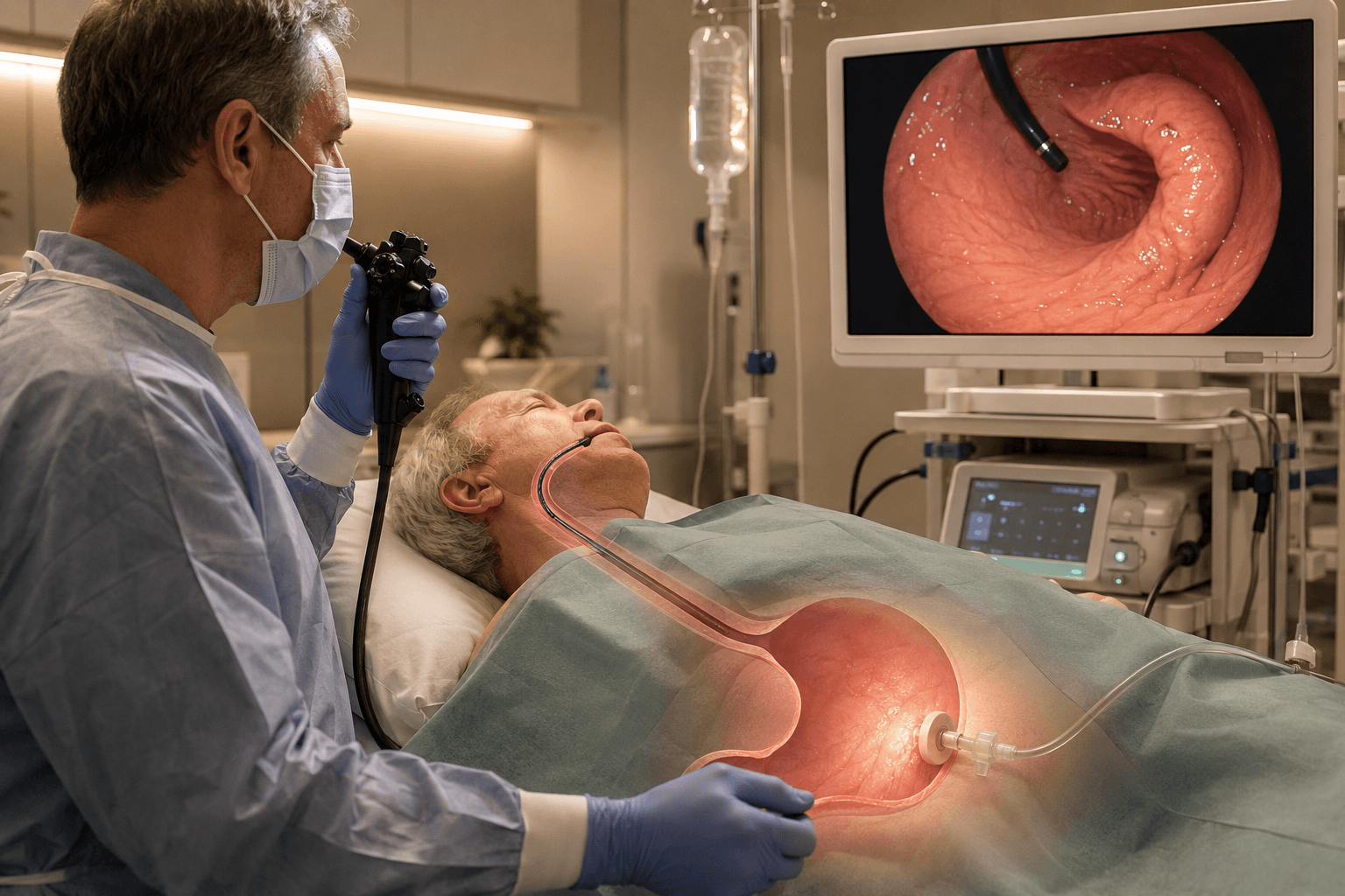
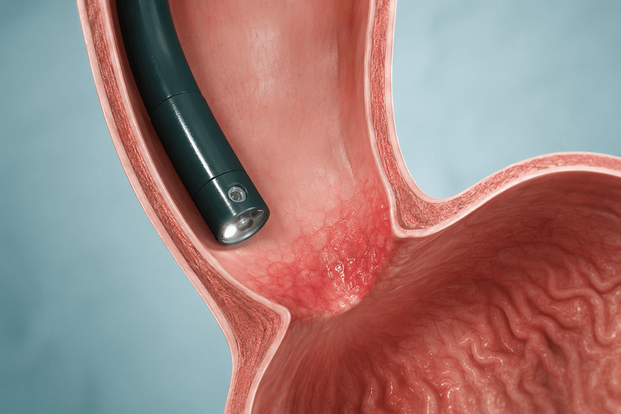
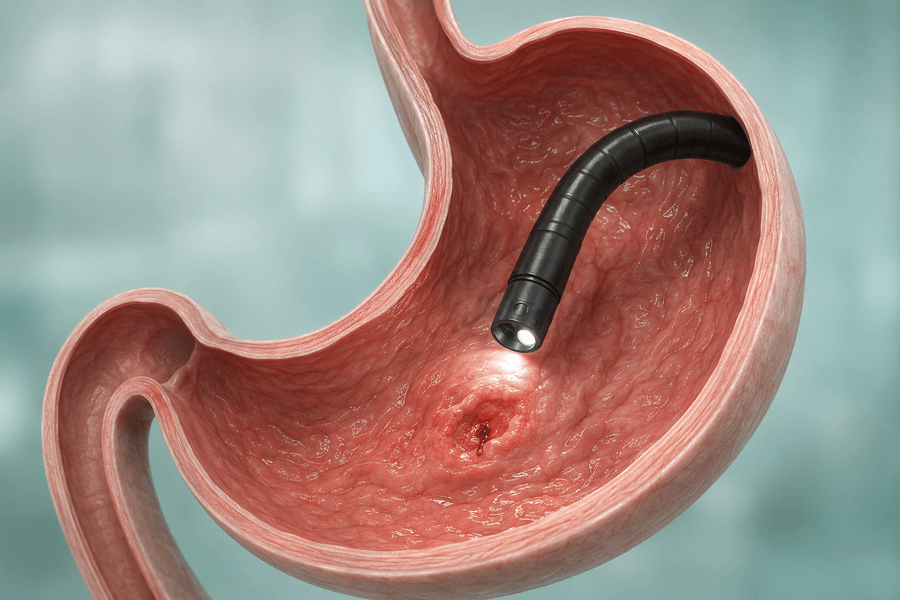
During the procedure, monitoring equipment is attached and sedation is generally used. Local anaesthetic numbs the abdominal skin. A flexible endoscope is passed through the mouth into the stomach, allowing the doctor to inspect the area and identify a safe placement site. A small opening is then made through the abdominal wall, and the tube is guided into position. Internal and external retaining devices help keep it secure.

The exact technique and time required depend on the patient’s anatomy and medical condition. Afterward, the patient is monitored until the sedation has worn off. The team confirms when water, medicines and feeding may begin. Current professional guidance supports early feeding in suitable patients, but the actual timing must follow the placing team’s instructions.

peg_tubes_procedure

Recovery and the First Few Days

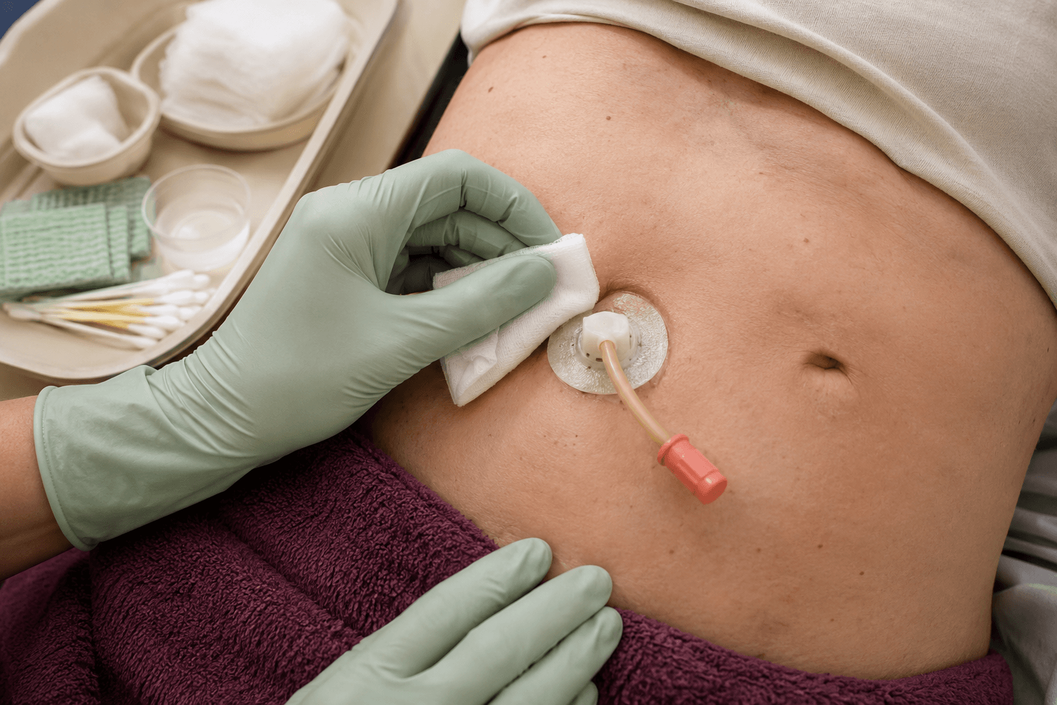
Tenderness around the site, mild abdominal discomfort or bloating can occur after insertion. The site should be observed for bleeding, leakage, increasing redness or swelling. Patients and caregivers should receive written instructions explaining how to clean the skin, secure the tube, flush it and give feeds or medicines.

Living Safely With a PEG Tube

Daily care helps protect the skin and keep the tube working. Patients should follow the instructions supplied by their own clinical team, as tube designs and care protocols differ. General principles include:

  • wash hands before handling the tube or preparing feed;
  • keep the stoma and surrounding skin clean and dry;
  • check the tube’s external position and securing device as instructed;
  • flush with the prescribed amount of water before and after feeds and medicines;
  • give medicines separately rather than mixing them into formula unless specifically advised;

peg_feeding_tubes_procedure

Possible Risks and Complications

PEG placement is commonly performed, but it is an invasive procedure and complications are possible. The individual risk depends on age, the underlying disease, nutritional status, medicines and other medical conditions. Potential problems include:

  • pain, bleeding or infection at the insertion site;
  • leakage of stomach contents and irritation of the surrounding skin;
  • tube blockage, damage, migration or accidental removal;
  • sedation-related breathing or cardiovascular problems;
  • aspiration of feed or stomach contents into the lungs;

The doctor will explain the most relevant risks before consent. The NHS patient information on PEG placement also emphasises fasting, antibiotic use in appropriate cases and discussion of procedural risks. Individual instructions from the treating team always take priority over general online guidance.

Warning Signs That Need Prompt Medical Attention

Contact the treating team promptly—or seek urgent medical care when symptoms are severe—if any of the following occur:

  • the tube falls out, moves significantly or appears longer or shorter than usual;
  • new or worsening abdominal pain, a rigid or swollen abdomen, or pain during feeding;
  • fever, chills, spreading redness, warmth, pus or an unpleasant smell around the site;
  • fresh bleeding, vomiting blood or black, tarry stools;
  • persistent leakage of feed or stomach contents around the tube;
  • repeated vomiting, severe diarrhoea or inability to tolerate feeds;

peg_feeding_tubes_patient_expectation

Can a PEG Tube Be Removed?

Yes. If swallowing improves and the person can again meet nutritional and hydration needs safely by mouth, the tube may no longer be necessary. The healthcare team should confirm this through medical, swallowing and dietetic review. Removal is performed by an appropriately trained clinician, and the opening usually closes afterward.

Some patients need replacement rather than removal because tubes wear, block or reach the recommended service interval. Routine follow-up allows the team to review the tube, skin, feeding tolerance, weight, hydration and ongoing need for enteral support.

Questions to Ask Before PEG Placement

  • What is the main goal of the PEG in this situation?
  • Is the need expected to be temporary or long term?
  • Are there reasonable alternatives, and what are their benefits and risks?
  • Can the patient continue any food or drink by mouth?
  • Who will create and monitor the nutrition plan?

peg_feeding_tubes_infographic

Conclusion

A PEG feeding tube can provide reliable access to nutrition, water and suitable medicines when swallowing is unsafe or oral intake is inadequate for an extended period. It may support recovery and help protect nutritional status, but it does not treat the underlying disease by itself.

If you or a family member has persistent swallowing difficulty, unintentional weight loss, repeated aspiration, or has been advised to consider longer-term tube feeding, contact Dr Eduan Deetlefs for an individual gastroenterology assessment. You can book an appointment with GI Doc or use the GI Doc contact page.

Dr Eduan Deetlefs Inc
Suite 304, 3rd Floor, The Park Building
Opposite Vincent Pallotti Hospital, Park Road
Pinelands, Cape Town
Telephone: 021 202 0626
Email: info@gidoc.co.za

GIDoc Cape Town

Patient-focused GI treatments and procedures in Cape Town.

Monday-Friday 8AM-4PM.

Connect with Us

© Dr. Eduan Deetlefs, Registered Gastroenterologist, GI Doc Cape Town

Our website information is not intended or implied to be a substitute for professional medical advice, diagnosis or treatment. Please consult a doctor about your specific condition. Only a trained physician can determine an accurate diagnosis and proper treatment.

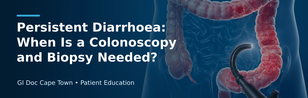
Persistent Diarrhoea: When Is a Colonoscopy and Biopsy Needed?

Persistent Diarrhoea: When Is a Colonoscopy and Biopsy Needed?

Persistent diarrhoea, unexplained weight loss and ongoing abdominal pain should not automatically be blamed on stress, food intolerance or a short-lived stomach infection. When loose or watery stools continue, recur frequently or occur with warning symptoms, they may signal inflammation, infection, medication-related injury or another digestive condition that needs proper investigation. A colonoscopy allows a gastroenterologist to inspect the colon directly, while biopsies can reveal microscopic disease that is invisible during the examination.

Diarrhoea is usually described as passing loose or watery stools more often than is normal for the individual. A brief episode is commonly caused by infection or a change in diet and often settles within several days. Persistent or chronic diarrhoea is different. Doctors generally use the term chronic diarrhoea when symptoms continue for four weeks or longer, although earlier assessment may be necessary when symptoms are severe or accompanied by red flags.

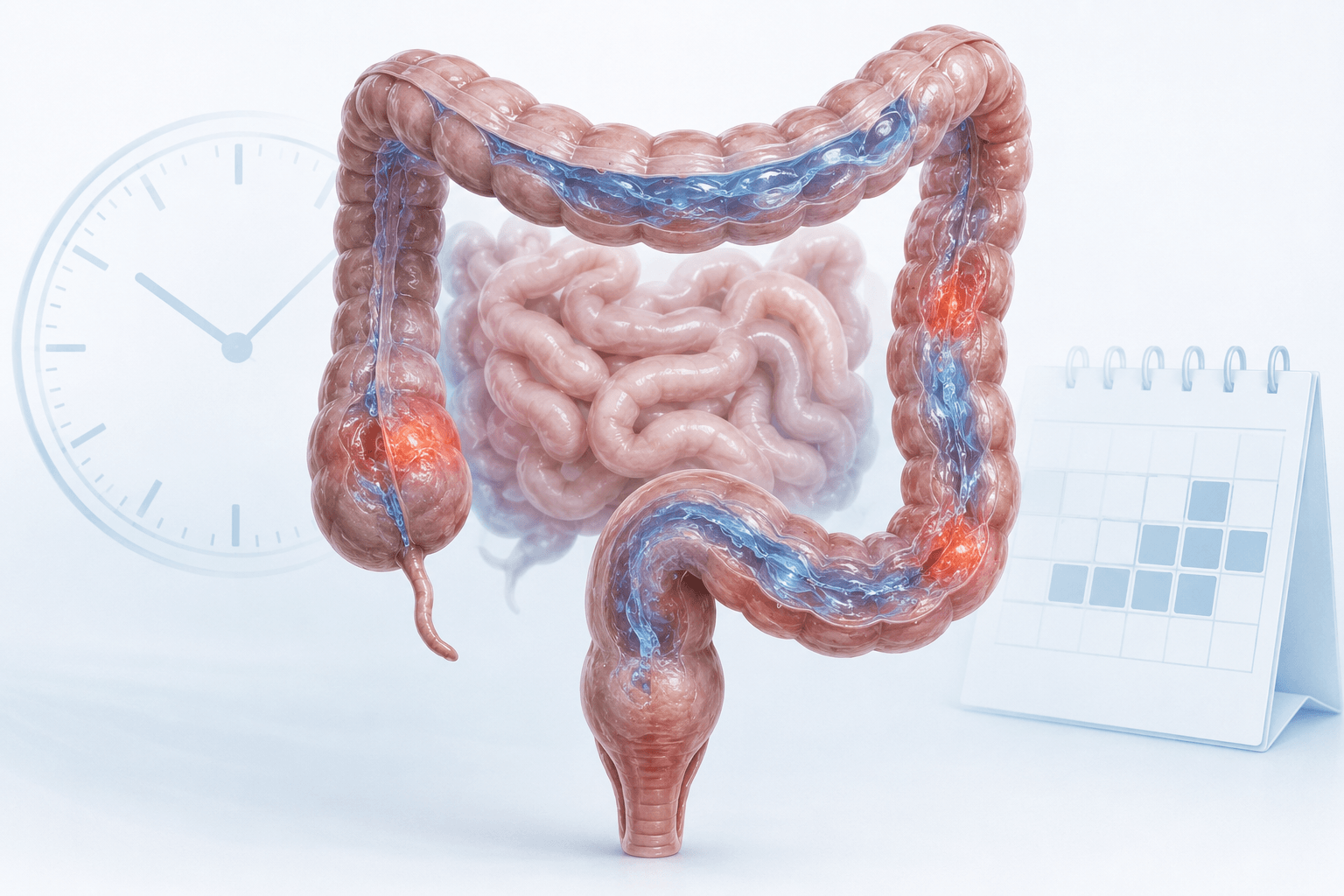
persistent_diarrhoea_inflammationPersistent diarrhoea may arise from inflammatory, infectious, medication-related or functional causes.

Why persistent diarrhoea needs a structured investigation

Diarrhoea is a symptom rather than a diagnosis. Different conditions can produce very similar bowel changes, so treatment based only on symptoms may delay the correct diagnosis. For example, irritable bowel syndrome with diarrhoea can cause urgency and loose stools without visible inflammation, while inflammatory bowel disease may produce diarrhoea through active inflammation and ulceration. Microscopic colitis may cause frequent watery diarrhoea even though the colon appears normal during colonoscopy.

A gastroenterologist begins by considering the pattern of symptoms, the patient’s age, recent travel, diet, medicines, family history and other health conditions. Important questions include:

  • How long have the symptoms been present?
  • Are stools watery, greasy, bloody or associated with mucus?
  • Does diarrhoea wake the patient at night?
  • Is there urgency, incontinence, fever, pain or weight loss?
  • Have antibiotics, anti-inflammatory pain medicines or other drugs been used?
  • Is there a family history of inflammatory bowel disease, coeliac disease or colorectal cancer?

These details help distinguish between possible infectious, inflammatory, malabsorptive, medication-related and functional causes.

What may cause diarrhoea that does not settle?

Persistent diarrhoea has many possible causes. Some can be identified with blood or stool tests, while others require imaging, endoscopy or biopsy.

Inflammatory bowel disease

Crohn’s disease and ulcerative colitis are the two main forms of inflammatory bowel disease (IBD). They may cause ongoing diarrhoea, abdominal pain, blood or mucus in the stool, fatigue, anaemia, fever and weight loss. Symptoms can vary, and not every patient has visible bleeding.

A colonoscopy helps the specialist examine the colon and the final part of the small intestine, identify inflammation or ulcers and collect tissue samples. Biopsy findings help confirm the diagnosis and distinguish Crohn’s disease from ulcerative colitis and other forms of colitis. Patients living with Crohn’s disease or ulcerative colitis may also find information about IBD research and clinical studies useful when discussing future treatment options with their specialist.

Microscopic colitis

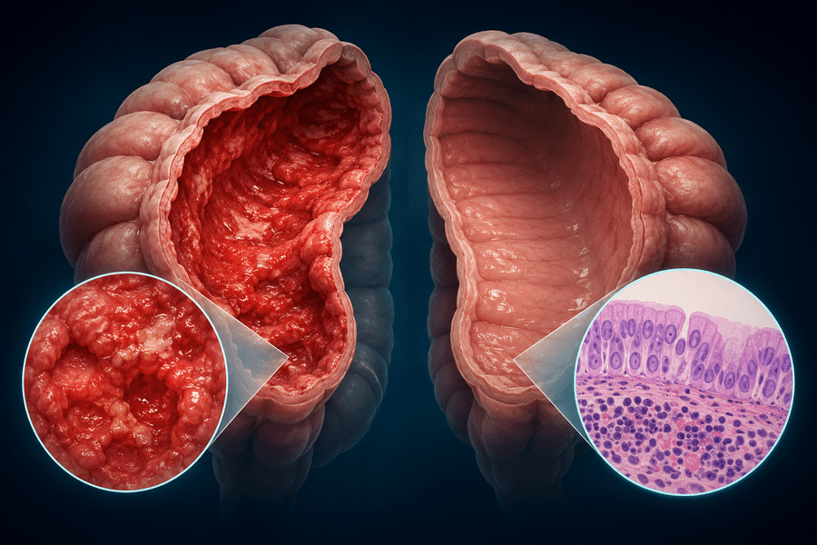
Microscopic colitis is an important cause of chronic, usually non-bloody watery diarrhoea. Its name reflects how it is diagnosed: the inflammation is seen under a microscope rather than necessarily being visible during colonoscopy. The colon lining may look healthy to the naked eye, so biopsies from appropriate areas of the colon are essential when the condition is suspected.

There are two main forms, collagenous colitis and lymphocytic colitis. Both can cause frequent watery stools, urgency, night-time diarrhoea, abdominal discomfort, fatigue and weight loss. The Mayo Clinic’s microscopic colitis guidance confirms that a colon tissue sample is needed because the bowel can appear normal during the examination.

Infection

Some infections last longer than the typical stomach bug, especially after travel, contaminated food or water, recent antibiotic use or in people with weakened immunity. Stool tests are usually performed before colonoscopy when an infectious cause is possible. This can include testing for bacteria, parasites or Clostridioides difficile. A colonoscopy is not the first test for most uncomplicated infections, but it may be considered when symptoms persist and routine testing does not provide an answer.

Coeliac disease and malabsorption

Coeliac disease is an immune reaction to gluten that damages the lining of the small intestine. It can cause diarrhoea, bloating, anaemia, nutrient deficiencies and weight loss. Blood tests are often used first. If results or symptoms suggest coeliac disease, a gastroscopy with duodenal biopsies may be recommended rather than colonoscopy alone.

Other problems involving digestion or absorption, including pancreatic disorders, bile acid diarrhoea and certain food intolerances, may require specialised tests. This is why the investigation should be tailored rather than relying on one procedure for every patient.

Medicines and supplements

Antibiotics, metformin, magnesium-containing products, laxatives, proton-pump inhibitors, some antidepressants and non-steroidal anti-inflammatory medicines can contribute to diarrhoea in certain people. Medicines may also be associated with microscopic colitis. Patients should provide a complete list of prescription medication, over-the-counter products and supplements, but should not stop prescribed treatment without medical advice.

Irritable bowel syndrome

Irritable bowel syndrome (IBS) can cause abdominal pain linked to bowel movements, bloating, urgency and altered stool consistency. However, IBS should not be assumed when warning signs are present. The Mayo Clinic’s IBS diagnostic guidance highlights red flags such as weight loss, rectal bleeding, fever, night-time diarrhoea and iron-deficiency anaemia, which may prompt further testing.

Which warning signs may make colonoscopy more urgent?

Not everyone with diarrhoea requires a colonoscopy. The decision depends on symptoms, examination findings, age, test results and individual risk. Colonoscopy is more likely to be recommended when diarrhoea is persistent or accompanied by:

  • Blood in the stool or black, tar-like stool
  • Unexplained weight loss
  • Iron-deficiency anaemia
  • Night-time symptoms that wake the patient
  • Ongoing or severe abdominal pain
  • Fever or signs of inflammation
  • A significant change in normal bowel habits
  • A family history of colorectal cancer, polyps or inflammatory bowel disease
  • Abnormal stool, blood or imaging results
  • New symptoms in an older adult
  • Symptoms that do not improve with appropriate initial management

The National Institute of Diabetes and Digestive and Kidney Diseases lists diarrhoea, rectal bleeding, abdominal pain and unexplained weight loss among the reasons doctors may use colonoscopy to investigate digestive symptoms.

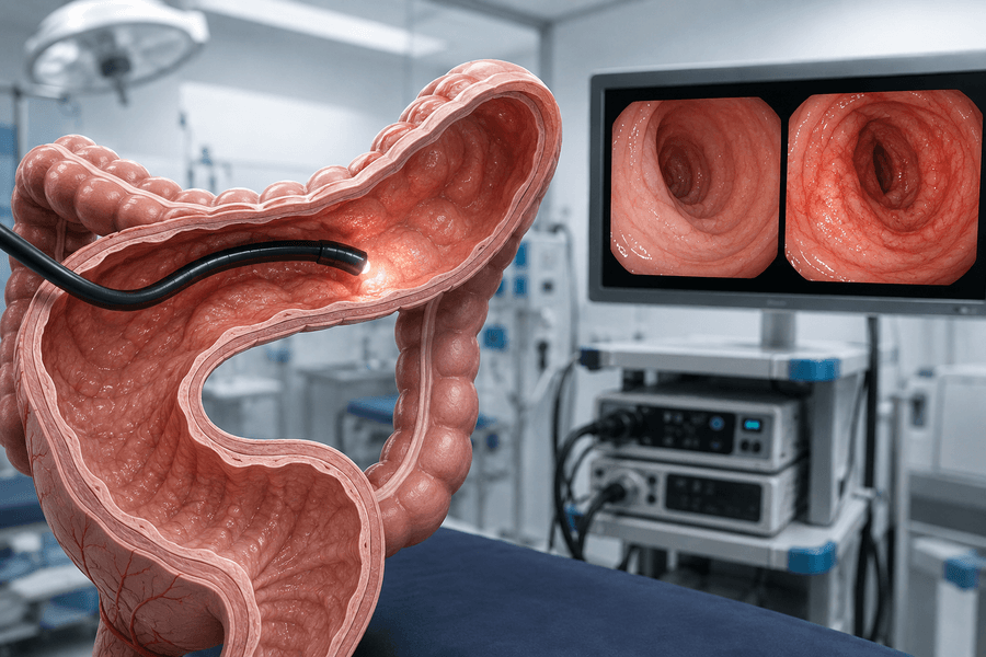
persistent_diarrhoea_colonoscopyColonoscopy provides a direct view of the colon and allows tissue samples to be collected.

What happens before colonoscopy is considered?

The initial assessment may include a physical examination and tests selected according to the patient’s history. Blood tests can look for anaemia, inflammation, electrolyte disturbance, thyroid disease or evidence of coeliac disease. Stool studies may check for infection, blood or markers of intestinal inflammation such as faecal calprotectin.

These tests help guide the next step, but normal results do not exclude every condition. In microscopic colitis, for example, the colon may look normal and inflammatory markers may be normal or only mildly raised. Persistent symptoms still deserve review when the clinical picture remains concerning.

Imaging may be useful when small-bowel disease, complications of Crohn’s disease, pancreatic disease or another structural problem is suspected. A capsule endoscopy or double-balloon enteroscopy may be considered when symptoms and results point beyond the reach of standard gastroscopy and colonoscopy.

What can colonoscopy detect?

During colonoscopy, a flexible camera is passed through the rectum to examine the lining of the colon. The procedure can identify:

  • Inflammation associated with ulcerative colitis or Crohn’s disease
  • Ulcers, erosions and bleeding sites
  • Colorectal polyps
  • Diverticular disease
  • Colorectal cancer
  • Some infectious or medication-related patterns of colitis
  • Narrowing or other structural abnormalities

The specialist may also examine the terminal ileum, which is the final part of the small intestine and a common site of Crohn’s disease. If a polyp or another suitable lesion is found, it may sometimes be removed during the same procedure. GI Doc provides a broader overview of its gastroenterology treatments and procedures, including diagnostic and therapeutic endoscopy.

Why take biopsies if the colon looks normal?

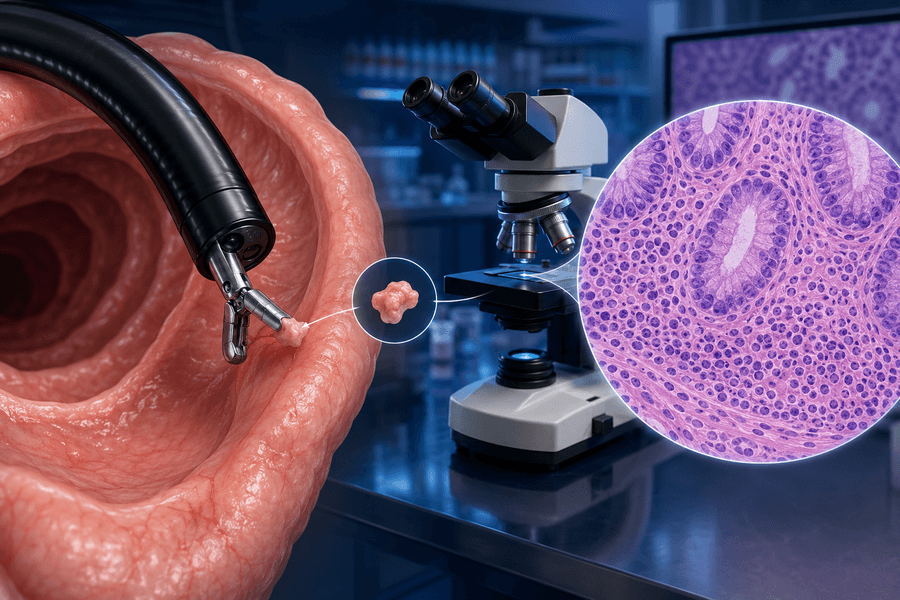
A biopsy is a very small tissue sample taken through the colonoscope. The patient does not usually feel it being collected. A pathologist then examines the sample under a microscope for inflammation, abnormal cells and other changes.

This step matters because a normal-looking colon does not always mean the tissue is normal. Microscopic colitis is the clearest example, but biopsies can also help assess early or treated IBD, infection and other forms of colitis. Samples may be taken from several sections because microscopic changes can be patchy.

biopsy_microscope_inflammationBiopsies may reveal inflammation that is not visible during colonoscopy.

A biopsy does not mean that cancer is suspected. It is a standard diagnostic tool that can provide information unavailable from the camera view alone. Patients awaiting pathology results should ask when and how these will be communicated, as the final diagnosis may depend on the laboratory report rather than the immediate visual findings.

Preparing for the procedure

The bowel needs to be clean so the specialist can inspect the lining thoroughly. Patients receive specific instructions about a clear-fluid diet and bowel-cleansing medication. Following these instructions closely improves visibility and reduces the chance that the examination will need to be repeated.

Before the procedure, patients should discuss all medicines, allergies and medical conditions with the team. Blood thinners and diabetes medication may require individual instructions. These medicines must not be changed without guidance from the prescribing clinician or endoscopy team.

Sedation is commonly used, so patients generally need someone to take them home. They should follow the practice’s instructions about driving, alcohol, work and important decisions after the procedure. The Mayo Clinic colonoscopy overview explains that colonoscopy is used both for screening and for investigating long-lasting diarrhoea, bleeding and other bowel symptoms.

What happens after colonoscopy?

Temporary bloating, gas or mild cramping can occur after the examination. The doctor may explain visible findings before discharge, while biopsy results usually take longer. Treatment depends on the diagnosis and may include medication, dietary management, correction of fluid or nutrient deficiencies, treatment of infection or a longer-term IBD care plan.

If Crohn’s disease or ulcerative colitis is diagnosed, treatment and monitoring are personalised according to the disease location, severity and response. Spoke Research explains how clinical research is advancing IBD care; trial participation is voluntary and should always be discussed with the treating specialist.

ibd_vs_microscopic_colitisIBD can cause visible inflammation, while microscopic colitis may require biopsy for diagnosis.

When to seek urgent care

Persistent diarrhoea can cause dehydration and disturbances in salts such as sodium and potassium. Seek prompt medical care for inability to keep fluids down, marked weakness, reduced urination, severe or worsening abdominal pain, high fever, significant rectal bleeding, black stools, fainting or confusion. These symptoms should not wait for a routine appointment.

After colonoscopy, urgent medical attention is required for severe or increasing abdominal pain, persistent vomiting, fever, heavy rectal bleeding, dizziness or collapse. Patients should follow the emergency instructions supplied by the treating facility.

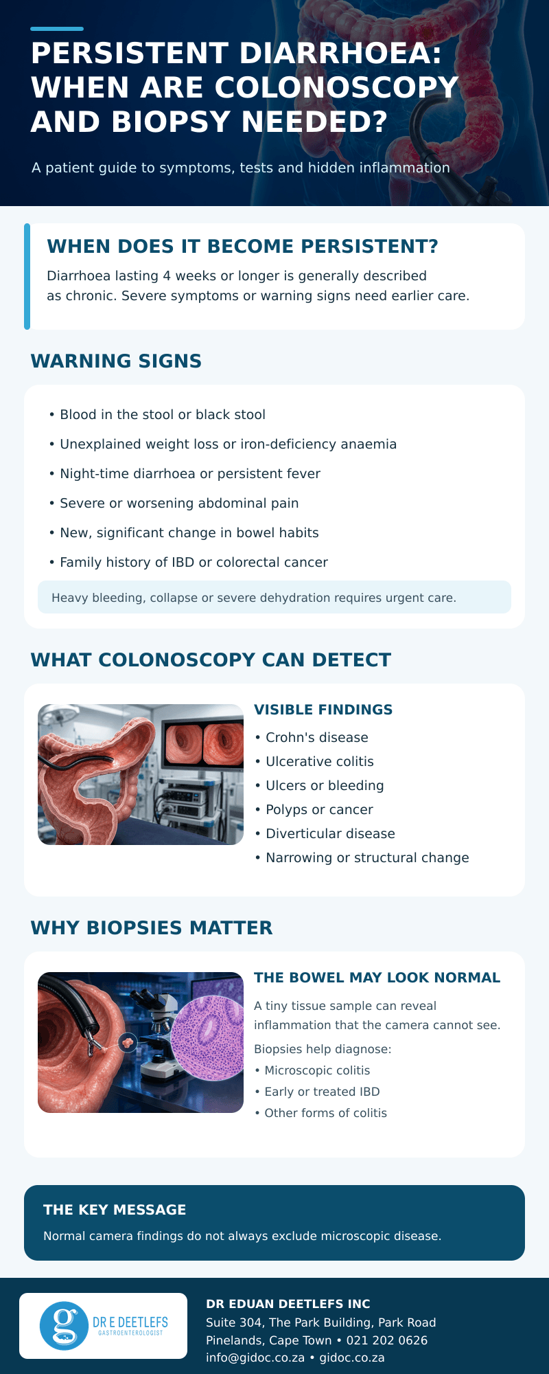
persistent_diarrhoea_infographic

Conclusion

Persistent diarrhoea should be assessed according to its duration, pattern and accompanying symptoms. Stool and blood tests may identify the cause in some patients, while others need colonoscopy to look for inflammation, ulcers, polyps, cancer or other structural disease. Biopsies are especially important because microscopic colitis and other tissue-level abnormalities may be present even when the colon looks normal.

If you have diarrhoea that is not settling, night-time symptoms, blood in the stool, unexplained weight loss, anaemia or ongoing abdominal pain, contact Dr Eduan Deetlefs, gastroenterologist in Cape Town, for an individual assessment.

Dr Eduan Deetlefs Inc: Suite 304, 3rd Floor, The Park Building, opposite Vincent Pallotti Hospital, Park Road, Pinelands, Cape Town, 7405. Telephone: 021 202 0626. Email: info@gidoc.co.za. Office hours: Monday to Thursday, 08:00-15:00; Friday is the in-room procedure day, 07:00-16:00.

This article is for general educational purposes and is not a substitute for individual medical advice, diagnosis or treatment.

GIDoc Cape Town

Patient-focused GI treatments and procedures in Cape Town.

Monday-Friday 8AM-4PM.

Connect with Us

© Dr. Eduan Deetlefs, Registered Gastroenterologist, GI Doc Cape Town

Our website information is not intended or implied to be a substitute for professional medical advice, diagnosis or treatment. Please consult a doctor about your specific condition. Only a trained physician can determine an accurate diagnosis and proper treatment.

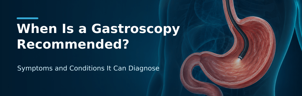
When Is a Gastroscopy Recommended? Symptoms and Conditions It Can Diagnose

When Is a Gastroscopy Recommended? Symptoms and Conditions It Can Diagnose


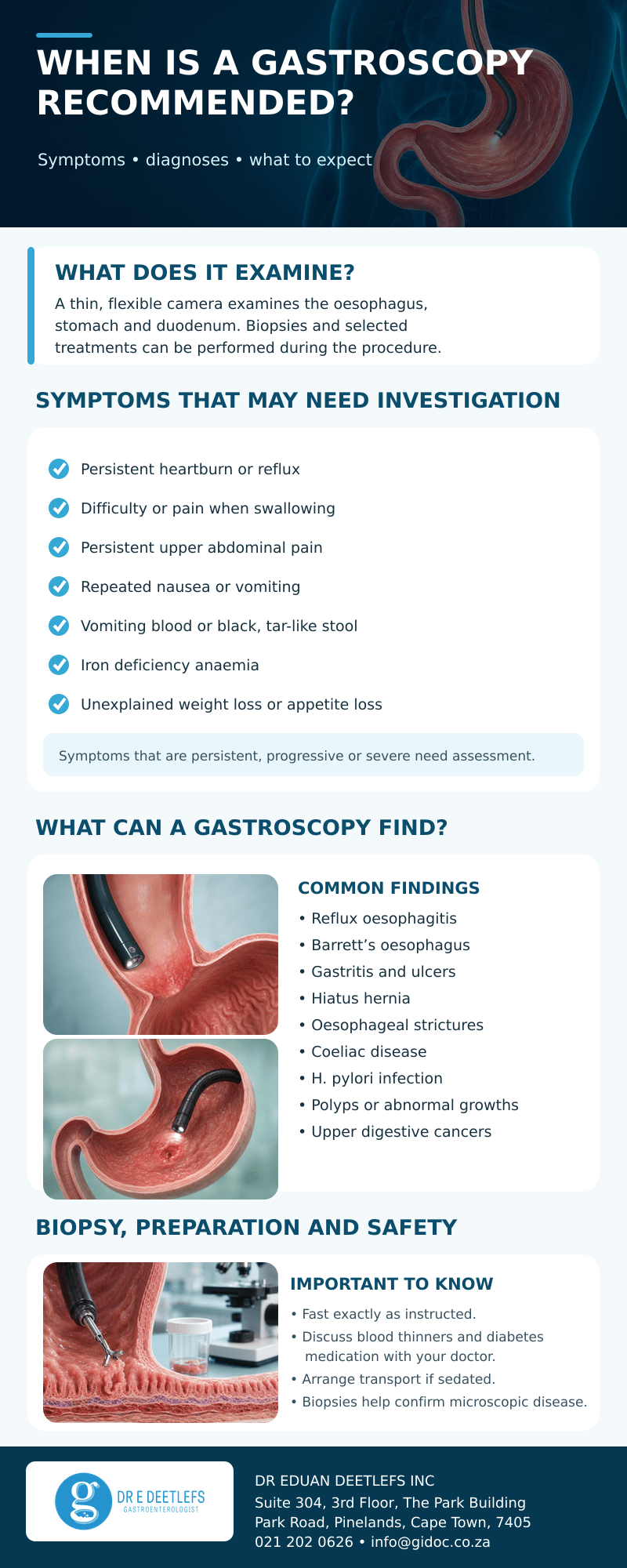
Persistent heartburn, dysphagia and unexplained bleeding should not be ignored. Although these symptoms do not always indicate a serious disease, they may arise from conditions affecting the oesophagus, stomach or first part of the small intestine. A gastroscopy allows a gastroenterologist to examine these areas directly, take biopsies when necessary and, in selected cases, provide treatment during the same procedure.

A gastroscopy is also called an upper gastrointestinal endoscopy or oesophagogastroduodenoscopy. It uses a thin, flexible instrument called an endoscope, which has a light and camera at its tip. The endoscope passes through the mouth, down the oesophagus and into the stomach and duodenum. Images are displayed on a monitor, allowing the specialist to assess the lining of the upper digestive tract in detail.

Gastroscopy_Upper_Digestive_Tract

A gastroscopy examines the oesophagus, stomach and duodenum.

When might a gastroscopy be recommended?

A gastroscopy is not required for every episode of indigestion or stomach discomfort. Short-lived symptoms often improve with simple treatment or lifestyle changes. However, further investigation may be appropriate when symptoms persist, recur, worsen, fail to respond to treatment or occur with warning signs.

The decision is based on the patient’s age, medical history, medicines, family history, examination and test results. A specialist may recommend gastroscopy for one or more of the following concerns.

Persistent heartburn or acid reflux

Occasional reflux is common, but frequent or persistent symptoms may require investigation, particularly when medication does not provide adequate relief. Gastroscopy can identify inflammation caused by reflux, ulceration, narrowing of the oesophagus or changes associated with Barrett’s oesophagus.

Barrett’s oesophagus occurs when long-term acid exposure changes the cells lining the lower oesophagus. It does not mean that cancer is present, but it may require biopsy and follow-up because it is associated with an increased risk of oesophageal cancer. GI Doc explains in more detail when acid reflux may warrant a gastroscopy.

Gastroscopy_Reflux_BarrettsGastroscopy can assess reflux-related inflammation and Barrett’s oesophagus.

Difficulty or pain when swallowing

The sensation that food sticks in the chest, swallowing that becomes progressively more difficult, or pain when swallowing needs medical assessment. These symptoms may be caused by inflammation, scarring, a stricture, an abnormal ring, eosinophilic oesophagitis, a motility disorder or, less commonly, a tumour.

Gastroscopy helps the specialist see whether a physical narrowing or abnormal area is present. Biopsies may be taken even when the lining looks relatively normal, because conditions such as eosinophilic oesophagitis can require microscopic confirmation. In suitable cases, a narrowed area may be treated with endoscopic dilatation.

Gastroscopy_Difficulty_SwallowingA narrowed oesophagus may contribute to difficulty swallowing.

Persistent upper abdominal pain or indigestion

Ongoing discomfort in the upper abdomen, recurrent indigestion, early fullness after eating or unexplained bloating may be linked to gastritis, peptic ulcer disease, infection or other upper digestive conditions. Symptoms alone cannot reliably distinguish one cause from another.

Gastroscopy gives a direct view of the stomach and duodenal lining. It can identify ulcers, erosions, inflammation and structural abnormalities such as a hiatus hernia. It is important to understand that some people with these symptoms have a normal gastroscopy. A normal examination is still useful because it helps exclude visible disease and guides the next stage of evaluation.

Recurrent nausea or vomiting

Persistent or unexplained nausea and vomiting may justify gastroscopy, especially when accompanied by weight loss, dehydration, pain, anaemia or difficulty eating. The procedure may detect inflammation, ulceration, narrowing or an obstruction at the stomach outlet. Other causes, including medication effects, metabolic disorders and delayed stomach emptying, may need different tests.

Signs of upper gastrointestinal bleeding

Vomiting fresh blood, vomiting material that resembles coffee grounds, or passing black, tar-like stool may indicate bleeding from the oesophagus, stomach or duodenum. These symptoms require prompt medical assessment. A gastroscopy can identify causes such as a bleeding ulcer, severe inflammation, a tear, enlarged veins called varices, or an abnormal growth.

Gastroscopy can be both diagnostic and therapeutic. Depending on the finding, a gastroenterologist may inject medication, apply a haemostatic clip or use another endoscopic technique to control bleeding. GI Doc offers both diagnostic and therapeutic gastroscopy and endoscopy procedures.

Gastroscopy_Ulcer_BleedingGastroscopy can identify ulcers and sources of upper gastrointestinal bleeding.

Iron deficiency anaemia

Iron deficiency anaemia can result from gradual blood loss that is not visible. When diet, menstrual loss and other common causes do not fully explain the problem, the digestive tract may need investigation. Gastroscopy may identify ulcers, inflammation, vascular lesions, coeliac disease or an upper digestive tumour. Depending on the patient’s age and circumstances, colonoscopy or small-bowel tests may also be recommended.

Unexplained weight loss or loss of appetite

Unintentional weight loss, reduced appetite, early satiety or progressive symptoms deserve medical attention. These features do not automatically mean cancer, but they are considered warning symptoms because they may occur with significant inflammation, ulcer disease, obstruction or malignancy. Direct examination and biopsy can help establish or exclude important causes.

Abnormal imaging or follow-up of a known condition

Gastroscopy may be advised after an abnormality is seen on a scan or contrast study. It may also be used to monitor a known condition, assess healing after treatment, investigate recurrent symptoms or follow a previously identified abnormality. The timing depends on the condition and the person’s individual risk.

What conditions can a gastroscopy diagnose?

According to the National Institute of Diabetes and Digestive and Kidney Diseases, upper GI endoscopy can help identify conditions affecting the oesophagus, stomach and duodenum. Findings may include:

  • Gastro-oesophageal reflux disease and reflux oesophagitis
  • Barrett’s oesophagus
  • Gastritis and duodenitis
  • Gastric or duodenal ulcers
  • Hiatus hernia
  • Oesophageal strictures, rings or other narrowing
  • Eosinophilic oesophagitis
  • Coeliac disease, when confirmed by duodenal biopsies
  • Helicobacter pylori-associated gastritis
  • Oesophageal or gastric varices
  • Benign polyps and other abnormal growths
  • Oesophageal, stomach or upper duodenal cancer

Not every condition is visible to the naked eye. This is why biopsy can be an important part of the examination.

Why are biopsies taken?

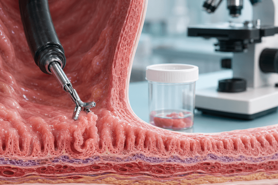
A biopsy is a tiny sample of tissue collected through the endoscope. Patients do not usually feel the tissue being taken. The sample is examined by a pathologist under a microscope.

Biopsies may be used to test for Helicobacter pylori, confirm coeliac disease, diagnose eosinophilic oesophagitis, evaluate inflammation, assess Barrett’s oesophagus or determine whether abnormal cells are benign, precancerous or cancerous. A biopsy does not necessarily mean that cancer is suspected; it is a routine diagnostic tool in many upper digestive conditions.

Gastroscopy_Biopsy
Small tissue samples can be collected for microscopic analysis.

What happens before and during the procedure?

Patients receive specific preparation instructions from their doctor. Fasting is necessary so the stomach is empty and the lining can be seen clearly. The Mayo Clinic’s upper endoscopy guidance also emphasises discussing medicines beforehand, especially blood thinners, diabetes treatment, iron supplements and anti-inflammatory pain medicines. Patients should not stop prescribed medication unless instructed by their healthcare professional.

Before the procedure, the team reviews the patient’s medical history, allergies and consent. A throat-numbing spray and/or sedation may be used. The patient lies on their side while the endoscope is gently passed through the mouth. It does not block breathing. The procedure itself is usually brief, although extra time may be needed if biopsies or treatment are performed.

Dr Deetlefs’ guide to what happens during a gastroscopy provides additional practical information for patients.

Afterwards, temporary bloating, mild nausea or a sore throat may occur. If sedation was given, the patient needs someone to take them home and must follow the practice’s restrictions on driving, alcohol, work and important decisions. Some findings may be discussed immediately, while biopsy results take longer.

Is gastroscopy safe?

Gastroscopy is generally considered a safe procedure. Complications are uncommon, but can include a reaction to sedation, bleeding, infection or a tear in the digestive tract. The risk may be higher when treatment is performed than when the procedure is purely diagnostic.

The NHS gastroscopy guidance advises urgent medical attention after the procedure for worsening chest or abdominal pain, breathing difficulty, fever, vomiting blood, black stool, or swallowing problems that are severe or getting worse. Patients should use the emergency contact instructions supplied by their treating facility.

Gastroscopy does not diagnose every digestive problem

Gastroscopy examines the lining of the upper digestive tract, but it does not assess the entire small bowel or colon. It also cannot by itself diagnose every functional, motility, pancreatic, gallbladder or liver disorder. Blood tests, ultrasound, CT or MRI, colonoscopy, capsule endoscopy, pH monitoring or other investigations may be more appropriate in some situations.

The aim is not simply to “have a scope,” but to select the investigation most likely to answer the clinical question. A gastroenterology consultation allows symptoms, risk factors and previous results to be considered together before a procedure is planned.

when_is_a_gastroscopy_recommended_infographic

Conclusion

A gastroscopy may be recommended when upper digestive symptoms are persistent, progressive or associated with warning signs such as difficulty swallowing, gastrointestinal bleeding, iron deficiency anaemia, repeated vomiting or unexplained weight loss. It can diagnose inflammation, ulcers, narrowing, Barrett’s oesophagus, coeliac disease and abnormal growths, while biopsy provides information that cannot always be seen during the examination. In some cases, treatment can also be performed during the procedure.

If you are experiencing these symptoms or have been advised to undergo a gastroscopy, contact Dr Eduan Deetlefs, gastroenterologist in Cape Town, for an assessment.

Dr Eduan Deetlefs Inc

Telephone: 021 202 0626
Email: info@gidoc.co.za
Website:  www.gidoc.co.za


GIDoc Cape Town

Patient-focused GI treatments and procedures in Cape Town.

Monday-Friday 8AM-4PM.

Connect with Us

© Dr. Eduan Deetlefs, Registered Gastroenterologist, GI Doc Cape Town

Our website information is not intended or implied to be a substitute for professional medical advice, diagnosis or treatment. Please consult a doctor about your specific condition. Only a trained physician can determine an accurate diagnosis and proper treatment.

Advanced Gastroenterology Treatments & Procedures at GI Doc Cape Town

Advanced Gastroenterology Treatments & Procedures at GI Doc Cape Town


Digestive symptoms are common, but they should not be ignored when they persist, recur, or begin to affect daily life. Persistent abdominal pain, rectal bleeding and unexplained weight loss can be warning signs of conditions that require specialist assessment. Other symptoms, including chronic heartburn, difficulty swallowing, bloating, prolonged diarrhoea, a change in bowel habits, jaundice or unexplained iron-deficiency anaemia, may also need investigation. Modern gastroenterology combines careful clinical assessment with advanced endoscopic procedures to identify the cause, treat selected problems and reduce the risk of complications.

At GI Doc Cape Town, Dr Eduan Deetlefs provides patient-focused care for gastrointestinal and liver-related conditions. He has a particular interest in inflammatory bowel disease (IBD) and iron-deficiency anaemia caused by obscure gastrointestinal bleeding. His services include diagnostic and therapeutic endoscopy, small-bowel investigation, colorectal cancer prevention and advanced interventional procedures.

Why Specialist Gastroenterology Care Matters


The digestive system includes the oesophagus, stomach, small intestine, colon, liver, gallbladder, pancreas and bile ducts. Because disorders in these organs can produce similar symptoms, the cause is not always obvious from symptoms alone. A gastroenterologist evaluates the full clinical picture, including the patient’s history, examination findings, blood tests, imaging and, where appropriate, endoscopy.

Early assessment is especially important when symptoms suggest gastrointestinal bleeding, bowel inflammation, obstruction or cancer. Procedures are selected according to the patient’s symptoms and risk factors; not every patient needs every test. In many cases, an endoscopic procedure can provide a diagnosis, obtain biopsies and deliver treatment during the same session.

gi_patient_care_gidoc_cpt

Gastroscopy for Upper Digestive Symptoms


A gastroscopy examines the oesophagus, stomach and duodenum using a thin, flexible camera passed through the mouth. It may be recommended for persistent reflux, upper abdominal pain, nausea or vomiting, difficulty swallowing, suspected ulcers, unexplained anaemia or signs of upper gastrointestinal bleeding.

The procedure can identify inflammation, ulcers, narrowing, coeliac disease, Barrett’s oesophagus and certain upper gastrointestinal cancers. Small tissue samples can be taken for laboratory analysis. Depending on the finding, treatment may also be performed, such as controlling a bleeding lesion or dilating a stricture. Patients with reflux-related cell changes may need surveillance or treatment because Barrett’s oesophagus can increase the risk of oesophageal cancer.

Colonoscopy, Cancer Prevention and Polypectomy


A colonoscopy allows the lining of the colon and rectum to be examined directly. It is used to investigate rectal bleeding, persistent diarrhoea, changes in bowel habits, unexplained anaemia and suspected IBD. It also plays an important role in colorectal cancer screening, even when a person has no symptoms.

One of the main advantages of colonoscopy is that polyps can often be removed during the examination. Many polyps are benign, but some types may become cancerous over time. A polypectomy interrupts this progression by removing abnormal tissue before it develops into cancer. Dr Deetlefs also performs endoscopic mucosal resection (EMR) for selected large, flat or complex lesions. These techniques may avoid more extensive surgery, although the safest approach depends on the lesion’s size, position and features.

advanced_gatroenterology_treatments_colonoscopy

Capsule Endoscopy and Double-Balloon Enteroscopy


The small intestine is difficult to examine fully with a standard gastroscopy or colonoscopy. Capsule endoscopy uses a swallowable camera to record images as it travels through the digestive tract. It is particularly useful when Crohn’s disease or a small-bowel source of blood loss is suspected after initial investigations have not found the cause. The test can identify ulcers, abnormal blood vessels and growths, but the capsule cannot take a biopsy or provide treatment. Patients with possible narrowing or obstruction need careful assessment because the capsule may become retained.

If capsule images reveal an abnormality, or if a deeper examination is needed, double-balloon enteroscopy can reach sections of the small bowel that conventional endoscopy cannot. It may be used to take biopsies, treat bleeding, remove selected polyps, or dilate a stricture. The United States National Institute of Diabetes and Digestive and Kidney Diseases provides further patient information about diagnosing gastrointestinal bleeding, including the role of capsule endoscopy.

advanced_treatments_deetlefs

ERCP for Bile Duct and Pancreatic Duct Disorders
 

Endoscopic retrograde cholangiopancreatography (ERCP) combines endoscopy with X-ray imaging to access the bile and pancreatic ducts. It is most often used when treatment is likely to be needed, for example to remove bile duct stones, relieve an obstruction, widen a narrowing or insert a stent.

Symptoms that may prompt investigation include jaundice, dark urine, pale stools, fever with upper abdominal pain or abnormal liver tests. ERCP can provide rapid relief when a duct is blocked, but it is a specialised procedure with recognised risks, including pancreatitis, bleeding, infection and perforation. Dr Deetlefs will assess whether ERCP is appropriate and explain the expected benefits, alternatives and risks.

Stricture Dilatation, Stents and Barrett’s Treatment


Inflammation, scarring, long-standing reflux or cancer can narrow part of the gastrointestinal tract. A stricture may cause food to stick, difficulty swallowing, vomiting, pain or signs of bowel obstruction. Endoscopic dilatation gently widens selected narrowed areas. In some situations, an oesophageal, gastric or duodenal stent may be placed to keep the passage open and improve swallowing or the movement of digestive contents.

For selected patients with Barrett’s oesophagus and abnormal cell changes, endoscopic mucosal resection or ablation may remove or destroy affected tissue. Ongoing reflux management and surveillance remain important because treatment and follow-up are tailored to the biopsy results and the degree of dysplasia.

advanced_gatroenterology_treatments_polypectomy

Specialist Care for Inflammatory Bowel Disease


Crohn’s disease and ulcerative colitis are chronic inflammatory conditions that may cause diarrhoea, bleeding, abdominal pain, fatigue, weight loss and nutritional deficiencies. Management may include endoscopy, imaging, laboratory monitoring, medicines, biological therapy and nutritional or iron support.

Dr Deetlefs provides ongoing IBD assessment and treatment planning, including an in-room infusion service for biological therapies and intravenous iron. He is also associated with Spoke Research Inc, a clinical trial centre focused on biological studies in IBD. The goal is not only to control symptoms, but also to monitor inflammation, prevent complications and support long-term quality of life.

When to Seek Prompt Medical Help


Arrange a medical assessment for persistent digestive symptoms, recurring bleeding, unexplained anaemia, difficulty swallowing or an ongoing change in bowel habits. Seek urgent medical care for vomiting blood, passing a large amount of blood, black tar-like stools, severe or worsening abdominal pain, a rigid or swollen abdomen, fainting, or jaundice accompanied by fever. Online information cannot replace an examination and an individual diagnosis.

advanced_gi_treatments_infographic

Conclusion


Advanced gastroenterology allows many digestive conditions to be investigated accurately and, in selected cases, treated without major surgery. The right procedure depends on the symptoms, medical history and findings from the initial consultation. Early specialist assessment can clarify the cause of ongoing symptoms, support timely treatment and reduce avoidable complications.

Patients in Cape Town who are experiencing persistent abdominal pain, reflux, rectal bleeding, difficulty swallowing, unexplained anaemia, weight loss or changes in bowel habits can contact Dr Eduan Deetlefs Inc to arrange an assessment.

Practice: Dr Eduan Deetlefs Inc | Telephone: 021 202 0626
Address: The Park Building (opposite Life Vincent Pallotti Hospital), Suite 304, 3rd Floor, Park Road, Pinelands, Cape Town, 7405
Email: info@gidoc.co.za
Website: www.gidoc.co.za

DISCLAIMER: PLEASE READ CAREFULLY

The information on this website is to provide general guidance. In no way does any of the information provided reflect definitive medical advice and self-diagnoses should not be made based on information obtained online. It is important to consult a Gastroenterologist or medical doctor regarding ANY and ALL symptoms or signs including, but not limited to: abdominal pain, haemorrhoids or anal / rectal bleeding as it may a sign of a serious illness or condition. A thorough consultation and examination should ALWAYS be performed for an accurate diagnosis and treatment plan. Be sure to call a physician or call our office today and schedule a consultation.

GIDoc Cape Town

Patient-focused GI treatments and procedures in Cape Town.

Monday-Friday 8AM-4PM.

Connect with Us

© Dr. Eduan Deetlefs, Registered Gastroenterologist, GI Doc Cape Town

Our website information is not intended or implied to be a substitute for professional medical advice, diagnosis or treatment. Please consult a doctor about your specific condition. Only a trained physician can determine an accurate diagnosis and proper treatment.

How Gastroenterologists Diagnose Chronic Abdominal Pain

How Gastroenterologists Diagnose Chronic Abdominal Pain

Abdominal pain is one of the most common reasons patients seek medical care. While occasional stomach discomfort may result from something as simple as indigestion or a mild stomach virus, chronic abdominal pain is a different matter entirely. When abdominal pain persists for weeks or months, it may indicate an underlying digestive condition that requires specialized evaluation and treatment.

Chronic abdominal pain, digestive disorders, and gastrointestinal health are closely linked, and identifying the root cause is often the first step toward lasting relief. Because many different conditions can produce similar symptoms, diagnosing chronic abdominal pain requires a comprehensive approach that combines medical history, physical examination, laboratory testing, and advanced diagnostic procedures.

At Dr. Eduan Deetlefs’ gastroenterology practice, patients benefit from a thorough diagnostic process designed to identify the cause of persistent abdominal discomfort and develop a personalized treatment plan.


What Is Chronic Abdominal Pain?

Chronic abdominal pain is generally defined as pain or discomfort occurring in the abdomen for three months or longer. The pain may be continuous or intermittent and can range from mild discomfort to severe symptoms that interfere with daily life.

Patients often describe chronic abdominal pain as:

  • Cramping
  • Burning
  • Sharp or stabbing pain
  • Dull aching
  • Bloating and pressure
  • Pain associated with eating
  • Pain associated with bowel movements

The location of the pain can also provide important clues. Pain in the upper abdomen may suggest stomach, esophageal, gallbladder, or pancreatic conditions, while lower abdominal pain may point toward intestinal disorders.

chronic_abdominal_pain_diagnose


Why Diagnosing Chronic Abdominal Pain Can Be Challenging

One of the biggest challenges in gastroenterology is that many digestive conditions produce overlapping symptoms.

For example, abdominal pain may be caused by:

Because symptoms often overlap, gastroenterologists must carefully investigate the source of pain rather than relying solely on symptom descriptions.

The First Step: A Detailed Medical History

The diagnostic process begins with a comprehensive discussion about the patient’s symptoms.

A gastroenterologist will typically ask:

  • When did the pain start?
  • Where is the pain located?
  • What does the pain feel like?
  • Does eating make it worse or better?
  • Are bowel habits changing?
  • Is there nausea or vomiting?
  • Is there unexplained weight loss?
  • Is there blood in the stool?
  • Is there a family history of digestive diseases?

This information helps narrow down potential causes and guides further testing.

Physical Examination

A physical examination remains an important part of diagnosing chronic abdominal pain.

During the examination, the doctor may:

  • Assess abdominal tenderness
  • Check for swelling or bloating
  • Listen to bowel sounds
  • Look for signs of inflammation
  • Evaluate for enlarged organs or masses

Certain findings can provide valuable clues that help determine which diagnostic tests should be performed next.

Laboratory Testing

Blood and stool tests are often among the first investigations ordered.

Common blood tests include:

Complete Blood Count (CBC)

A CBC can detect:

  • Infection
  • Inflammation
  • Anemia
  • Blood loss

Liver Function Tests

These tests evaluate whether liver or gallbladder problems may be contributing to symptoms.

Inflammatory Markers

Markers such as CRP and ESR can help identify inflammatory conditions like Crohn’s disease or ulcerative colitis.

Celiac Disease Screening

Specific blood tests can detect antibodies associated with celiac disease.

Stool Testing

Stool samples may be analyzed for:

  • Infection
  • Inflammation
  • Occult blood
  • Digestive abnormalities

These tests help eliminate many possible causes before more invasive investigations are considered.

chronic_abdominal_pain_gastroenterologist


Imaging Studies

Imaging plays a crucial role in diagnosing chronic abdominal pain.


Ultrasound

Ultrasound is commonly used to evaluate:

  • Gallstones
  • Liver disease
  • Kidney abnormalities
  • Certain abdominal masses

Because ultrasound does not involve radiation, it is often one of the first imaging tests performed.

CT Scan

Computed tomography (CT) scans provide detailed cross-sectional images of the abdomen and pelvis.

CT scans can help identify:

  • Inflammation
  • Tumors
  • Diverticulitis
  • Bowel obstruction
  • Abscesses

MRI

Magnetic Resonance Imaging (MRI) offers highly detailed imaging and is particularly useful in evaluating inflammatory bowel disease and certain liver disorders.


Endoscopy: Looking Directly Inside the Digestive Tract

One of the most valuable tools available to gastroenterologists is endoscopy.

Endoscopic procedures allow doctors to directly examine the lining of the digestive tract and identify abnormalities that may not appear on imaging studies.

Gastroscopy

A gastroscopy involves passing a thin flexible camera through the mouth into the esophagus, stomach, and upper small intestine.

Gastroscopy can diagnose:

  • Gastritis
  • Peptic ulcers
  • GERD
  • Hiatal hernias
  • Barrett’s esophagus
  • Stomach cancer

Biopsies can also be taken during the procedure for further analysis.

Colonoscopy

A colonoscopy examines the large intestine and rectum.

This procedure can detect:

  • Colon polyps
  • Colorectal cancer
  • Crohn’s disease
  • Ulcerative colitis
  • Diverticular disease

For patients with unexplained lower abdominal pain, colonoscopy often provides critical diagnostic information.

chronic_abdominal_pain_endoscopy_colonoscopy


Advanced Functional Testing

Sometimes standard tests fail to reveal the cause of chronic abdominal pain.

In these situations, advanced functional testing may be required.

pH Monitoring

This test measures acid exposure in the esophagus and helps diagnose GERD.

Esophageal Manometry

Manometry evaluates muscle contractions within the esophagus and assesses the function of the lower esophageal sphincter.

Breath Testing

Breath tests can help diagnose:

  • Lactose intolerance
  • Fructose intolerance
  • Small Intestinal Bacterial Overgrowth (SIBO)

These tests are particularly useful when symptoms include bloating, gas, and abdominal discomfort.

When Biopsies Are Necessary


In some cases, tissue samples must be collected to establish an accurate diagnosis.

Biopsies can help diagnose:

  • Celiac disease
  • Microscopic colitis
  • Barrett’s esophagus
  • Stomach infections
  • Gastrointestinal cancers

Biopsies are usually painless and can often be performed during endoscopy or colonoscopy procedures.

chronic_abdominal_pain_care


Red Flag Symptoms That Require Urgent Investigation

While many causes of abdominal pain are not life-threatening, some symptoms warrant immediate medical evaluation.

These include:

  • Unexplained weight loss
  • Persistent vomiting
  • Difficulty swallowing
  • Blood in the stool
  • Black stools
  • Severe anemia
  • Night-time symptoms
  • Family history of gastrointestinal cancer

The presence of these symptoms often prompts more urgent testing.

How Gastroenterologists Develop a Treatment Plan

Once the underlying cause is identified, treatment can begin.

Depending on the diagnosis, treatment may include:

  • Dietary modifications
  • Medication
  • Endoscopic procedures
  • Lifestyle changes
  • Surgical intervention when necessary

The goal is not simply to relieve symptoms but to address the root cause of the condition and prevent complications.

Why Early Diagnosis Matters

Many digestive conditions become easier to manage when diagnosed early.

Prompt diagnosis can:

  • Prevent complications
  • Improve quality of life
  • Reduce chronic pain
  • Identify serious diseases early
  • Improve treatment outcomes

Patients who delay seeking care may unknowingly allow underlying conditions to progress.

chronic_abdominal_care_info


Conclusion

Chronic abdominal pain is not a condition that should be ignored or simply managed with over-the-counter remedies. Because numerous digestive disorders can cause similar symptoms, accurate diagnosis requires specialized evaluation by an experienced gastroenterologist. Through a combination of medical history, physical examination, laboratory testing, imaging studies, endoscopy, and advanced functional assessments, gastroenterologists can identify the root cause of persistent abdominal discomfort and develop effective treatment strategies.

At Dr. Eduan Deetlefs’ gastroenterology practice, patients receive comprehensive diagnostic evaluations and evidence-based treatment plans tailored to their individual needs. Whether symptoms are caused by GERD, inflammatory bowel disease, ulcers, food intolerances, or other gastrointestinal conditions, early diagnosis is essential for achieving the best possible outcomes.

If you are experiencing ongoing abdominal pain, bloating, digestive discomfort, or unexplained gastrointestinal symptoms, contact Dr. Eduan Deetlefs to schedule a consultation.

Expert evaluation and timely treatment can help restore your digestive health and improve your quality of life.


GIDoc Cape Town

Patient-focused GI treatments and procedures in Cape Town.

Monday-Friday 8AM-4PM.

Connect with Us

© Dr. Eduan Deetlefs, Registered Gastroenterologist, GI Doc Cape Town

Our website information is not intended or implied to be a substitute for professional medical advice, diagnosis or treatment. Please consult a doctor about your specific condition. Only a trained physician can determine an accurate diagnosis and proper treatment.