021 202 0626
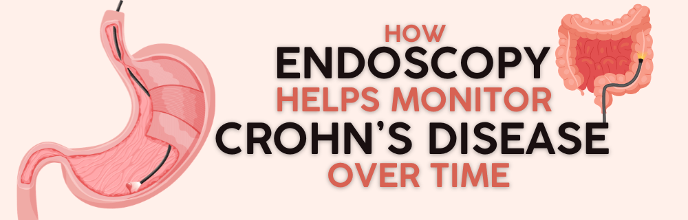
How Endoscopy Helps Monitor Crohn’s Disease Over Time

How Endoscopy Helps Monitor Crohn’s Disease Over Time


Crohn’s disease, endoscopy, and long-term monitoring are essential components in effectively managing this chronic and often unpredictable inflammatory bowel disease (IBD). While many patients focus on symptoms such as abdominal pain, diarrhoea, fatigue, and weight loss, these do not always reflect what is happening inside the digestive tract.

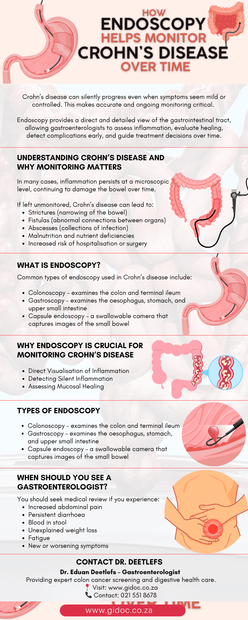
Crohn’s disease can silently progress even when symptoms seem mild or controlled. This makes accurate and ongoing monitoring critical. Endoscopy provides a direct and detailed view of the gastrointestinal tract, allowing gastroenterologists to assess inflammation, evaluate healing, detect complications early, and guide treatment decisions over time.

For patients under the care of specialists such as Dr. Deetlefs, endoscopy is not just a diagnostic tool—it is a cornerstone of long-term disease management aimed at achieving sustained remission and preventing complications.


Understanding Crohn’s Disease and Why Monitoring Matters


Crohn’s disease is a chronic inflammatory condition that can affect any part of the gastrointestinal tract, although it most commonly involves the small intestine and colon. It is characterised by an abnormal immune response that leads to ongoing inflammation of the bowel lining.

The disease typically follows a relapsing-remitting pattern:

  • Flare-ups: Periods of active inflammation and symptoms
  • Remission: Periods where symptoms improve or disappear

However, symptom relief does not always mean the disease is under control. In many cases, inflammation persists at a microscopic level, continuing to damage the bowel over time.

If left unmonitored, Crohn’s disease can lead to:

  • Strictures (narrowing of the bowel)
  • Fistulas (abnormal connections between organs)
  • Abscesses (collections of infection)
  • Malnutrition and nutrient deficiencies
  • Increased risk of hospitalisation or surgery

This is why regular monitoring—particularly through endoscopy—is essential. 


What Is Endoscopy?


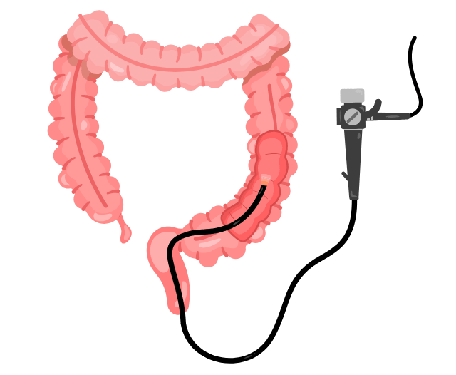
Endoscopy refers to a group of procedures that use a thin, flexible tube with a camera and light source to examine the inside of the digestive tract in real time.

Common types of endoscopy used in Crohn’s disease include:

  • Colonoscopy – examines the colon and terminal ileum
  • Gastroscopy (upper endoscopy) – examines the oesophagus, stomach, and upper small intestine
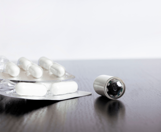
  • Capsule endoscopy – a swallowable camera that captures images of the small bowel

These procedures allow doctors to directly visualise inflammation, assess disease severity, and obtain tissue samples (biopsies) for further analysis.


colonoscopy_crohns_diagnose


Why Endoscopy Is Crucial for Monitoring Crohn’s Disease


Endoscopy provides insights that cannot be obtained through symptoms or blood tests alone.

  1. Direct Visualisation of Inflammation

Endoscopy allows gastroenterologists to see:

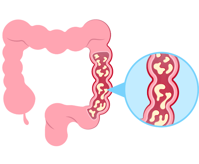
  • Redness and swelling of the bowel lining
  • Ulcers and erosions
  • Bleeding areas
  • Patterns of inflammation

This direct visual evidence helps determine how active the disease is and where it is located.

  1. Detecting Silent Inflammation

A key challenge in Crohn’s disease is that symptoms do not always match disease activity.

Patients may feel relatively well but still have ongoing inflammation that can lead to complications. This is known as subclinical or silent inflammation.

Endoscopy helps detect this early, allowing treatment to be adjusted before damage progresses.

  1. Assessing Mucosal Healing

Modern Crohn’s disease management focuses on achieving mucosal healing, where the bowel lining appears normal on endoscopy.

Mucosal healing is associated with:

  • Fewer disease flare-ups
  • Reduced need for hospitalisation
  • Lower risk of surgery
  • Improved long-term outcomes

Endoscopy is currently the most reliable method to confirm mucosal healing.


endoscopy_narrowing_crohns


When Is Endoscopy Recommended?


Endoscopy is used at different stages of Crohn’s disease:

At Initial Diagnosis

To confirm Crohn’s disease and determine its severity, extent, and location.

After Starting Treatment

To assess how well medications are controlling inflammation.

During Remission

To confirm that inflammation has resolved and healing has occurred.

When Symptoms Change or Worsen

To identify flare-ups or complications such as strictures or fistulas.

The frequency of endoscopy varies depending on the patient’s condition, but regular monitoring is essential for optimal disease control.


Types of Endoscopy Used in Crohn’s Disease Monitoring


Colonoscopy

Colonoscopy remains the gold standard for monitoring Crohn’s disease affecting the colon and terminal ileum.

It allows doctors to:

  • Examine the bowel lining in detail
  • Identify ulcers and inflammation
  • Detect narrowing or scarring
  • Take biopsies for microscopic evaluation

IBS vs IBD relevance:

While IBS shows a normal colon, Crohn’s disease reveals clear signs of inflammation or structural damage.


Gastroscopy (Upper Endoscopy)

Although less common, Crohn’s disease can affect the upper gastrointestinal tract.

Gastroscopy helps:

  • Detect inflammation in the stomach or duodenum
  • Rule out other conditions such as ulcers or gastritis
  • Provide a complete assessment of the digestive system

Capsule Endoscopy

Capsule endoscopy is particularly valuable for examining the small intestine, which is difficult to reach with traditional scopes.

Advantages include:

  • Non-invasive procedure
  • Comprehensive imaging of the small bowel
  • Detection of early or subtle disease

It is especially useful when symptoms persist but colonoscopy results are inconclusive.
endoscopy_crohns_disease

How Endoscopy Guides Treatment Decisions


Endoscopy plays a central role in the “treat-to-target” approach, where treatment is adjusted based on objective findings rather than symptoms alone.

If inflammation is still present:

  • Medication doses may be increased
  • Biologic therapies may be introduced or changed
  • Additional investigations may be required

If mucosal healing is achieved:

  • Current treatment may be continued
  • Monitoring intervals may be extended
  • Risk of complications is significantly reduced

This personalised approach ensures patients receive the most effective therapy for their disease stage.


Preventing Complications Through Regular Endoscopy


Crohn’s disease can lead to serious complications if not properly monitored.

Endoscopy helps detect:

  • Strictures: Narrowing of the bowel that can cause obstruction
  • Fistulas: Abnormal connections between organs
  • Abscesses: Localised infections
  • Pre-cancerous changes: Especially in long-standing disease

Early detection allows for timely intervention, reducing the likelihood of surgery and improving overall outcomes.


Advanced Endoscopic Treatments Offered by Dr. Deetlefs


Endoscopy is not only diagnostic—it can also be therapeutic.

Dr. Deetlefs offers advanced endoscopic procedures such as:

Endoscopic Dilatation of Strictures

  • Used to widen narrowed sections of the bowel
  • Helps relieve obstruction symptoms
  • May delay or avoid the need for surgery

Therapeutic Colonoscopy

  • Management of complications
  • Biopsy and targeted treatment
  • Ongoing surveillance of disease progression

These minimally invasive techniques are a key part of modern Crohn’s disease care.
crohns_disease_monitoring

The Role of Endoscopy in Long-Term Disease Control


Crohn’s disease is a lifelong condition, and long-term monitoring is essential for maintaining control.

Endoscopy helps:

  • Track disease progression over time
  • Evaluate treatment effectiveness
  • Ensure mucosal healing is achieved
  • Detect complications early

It is often used alongside:

  • Blood tests (CRP, ESR)
  • Stool tests (fecal calprotectin)
  • Imaging (MRI or CT scans)

Together, these tools provide a comprehensive understanding of disease activity.


Are There Risks Associated with Endoscopy?


Endoscopy is generally safe and well-tolerated.

Potential risks (rare):

  • Bleeding after biopsy
  • Perforation of the bowel
  • Reaction to sedation

Your gastroenterologist will carefully assess your suitability and explain the procedure beforehand. In most cases, the benefits of accurate monitoring far outweigh the risks.


When Should You See a Gastroenterologist?


If you have Crohn’s disease, ongoing specialist care is essential—even during remission.

You should seek medical review if you experience:

  • Increased abdominal pain
  • Persistent diarrhoea
  • Blood in stool
  • Unexplained weight loss
  • Fatigue
  • New or worsening symptoms

Regular follow-up and endoscopic monitoring ensure early intervention and better long-term outcomes.
how_endoscopy_helps_monitor_crohns_infographic


Conclusion


Endoscopy plays a critical role in monitoring Crohn’s disease over time. By providing direct visualisation of the gastrointestinal tract, it allows gastroenterologists to detect inflammation, confirm healing, and identify complications before they become severe.

When combined with a personalised treatment plan, regular endoscopic monitoring helps patients achieve long-term remission, reduce flare-ups, and improve quality of life.

Under the expert care of a gastroenterologist such as Dr. Deetlefs, endoscopy becomes an essential tool in managing Crohn’s disease effectively and proactively.


GIDoc Cape Town

Patient-focused GI treatments and procedures in Cape Town.

Monday-Friday 8AM-4PM.

Connect with Us

© Dr. Eduan Deetlefs, Registered Gastroenterologist, GI Doc Cape Town

Our website information is not intended or implied to be a substitute for professional medical advice, diagnosis or treatment. Please consult a doctor about your specific condition. Only a trained physician can determine an accurate diagnosis and proper treatment.
What Tests Are Used to Diagnose IBS vs IBD?

What Tests Are Used to Diagnose IBS vs IBD?


Irritable Bowel Syndrome (IBS), Inflammatory Bowel Disease (IBD), and gastrointestinal diagnostic testing are among the most frequently searched digestive health concerns worldwide. Patients often experience ongoing symptoms such as abdominal pain, bloating, diarrhoea, constipation, fatigue, and urgency — yet the underlying cause can vary significantly.

Distinguishing IBS from IBD is essential because the two conditions differ fundamentally in pathology, severity, and treatment approach. IBS is a functional gastrointestinal disorder, meaning the bowel looks structurally normal but does not function properly.

IBD, which includes Crohn’s disease and ulcerative colitis, is a chronic inflammatory autoimmune condition that causes visible damage to the digestive tract.

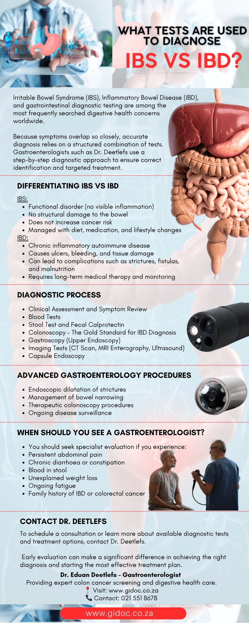
Because symptoms overlap so closely, accurate diagnosis relies on a structured combination of clinical evaluation, laboratory testing, stool analysis, imaging, and endoscopic procedures. Gastroenterologists such as Dr. Deetlefs use a step-by-step diagnostic approach to ensure correct identification and targeted treatment.


Why Differentiating IBS vs IBD Is So Important


Although IBS and IBD share similar symptoms, they have very different implications for long-term health.

IBS:

  • Functional disorder (no visible inflammation)
  • No structural damage to the bowel
  • Does not increase cancer risk
  • Managed with diet, medication, and lifestyle changes

IBD:

  • Chronic inflammatory autoimmune disease
  • Causes ulcers, bleeding, and tissue damage
  • Can lead to complications such as strictures, fistulas, and malnutrition
  • Requires long-term medical therapy and monitoring

Because of these differences, misdiagnosis can delay appropriate treatment and allow inflammation in IBD to progress unchecked. This is why a structured diagnostic process is essential.

ibs_vs_ibd

Step 1: Clinical Assessment and Symptom Review


The diagnostic process always begins with a detailed consultation.

A gastroenterologist will assess:

  • Nature and duration of symptoms
  • Stool patterns (diarrhoea, constipation, or alternating)
  • Presence of blood or mucus in stool
  • Weight loss or fatigue
  • Family history of IBD, celiac disease, or colorectal cancer
  • Trigger factors such as stress or food

IBS symptoms often fluctuate and are strongly linked to stress and diet. IBD symptoms tend to be more persistent and progressive.

However, symptoms alone are not sufficient for diagnosis, which is why further testing is required.


Step 2: Blood Tests for IBS vs IBD


Blood tests are an essential first-line diagnostic tool.

Common blood tests include:

  • Full blood count (FBC)
  • C-reactive protein (CRP)
  • Erythrocyte sedimentation rate (ESR)
  • Iron studies (ferritin, iron, transferrin)
  • Vitamin B12 and folate levels
  • Liver function tests

What results typically show:

In IBS:

  • Blood tests are usually completely normal
  • No signs of inflammation or deficiency

In IBD:

  • Elevated CRP and ESR indicate inflammation
  • Anemia may be present due to chronic blood loss
  • Vitamin deficiencies are common due to malabsorption

These markers help differentiate functional bowel disorders from inflammatory disease, but they are not definitive on their own.

Step 3: Stool Tests and Fecal Calprotectin


Stool testing is one of the most powerful non-invasive tools in differentiating IBS from IBD.

Key stool tests include:

  • Fecal calprotectin
  • Stool culture (infection screening)
  • Stool microscopy for parasites
  • Occult blood test (hidden blood detection)

Fecal calprotectin explained:

Calprotectin is a protein released by white blood cells during intestinal inflammation. Measuring its level provides a direct indication of inflammation in the bowel.

  • Low calprotectin: Likely IBS or functional disorder
  • High calprotectin: Strongly suggests IBD
  • Borderline results: Require further investigation

This test is extremely useful because it helps avoid unnecessary colonoscopies in low-risk patients while identifying those who require urgent investigation.

Step 4: Colonoscopy – The Gold Standard for IBD Diagnosis


A colonoscopy is one of the most important diagnostic procedures in gastroenterology and is central to diagnosing IBD.

What happens during a colonoscopy:

  • A flexible camera is inserted into the rectum
  • The entire colon is visually examined
  • Any inflammation, ulcers, or bleeding is recorded
  • Tissue biopsies are taken for histological analysis

Conditions diagnosed include:

IBS vs IBD findings:

  • IBS: Normal-looking colon with no inflammation
  • IBD: Visible inflammation, ulceration, or structural changes

Biopsy results confirm the presence and type of inflammatory disease, making this test critical for diagnosis.


ibs_vs_ibd_diagnose


Step 5: Gastroscopy (Upper Endoscopy)


A gastroscopy may be performed when symptoms extend to the upper digestive tract.

It evaluates:

  • Oesophagus
  • Stomach
  • Duodenum (upper small intestine)

It helps diagnose or exclude:

  • Celiac disease
  • Gastritis
  • Peptic ulcers
  • Helicobacter pylori infection

While not directly used to diagnose IBS, it helps rule out other causes of digestive symptoms.


Step 6: Imaging Tests (CT Scan, MRI Enterography, Ultrasound)


When Crohn’s disease is suspected, especially in the small intestine, imaging plays a key role.

Imaging methods:

  • CT enterography
  • MRI enterography (preferred for younger patients or long-term monitoring)
  • Abdominal ultrasound (in selected cases)

What imaging detects:

  • Bowel wall thickening
  • Deep tissue inflammation
  • Abscesses
  • Fistulas
  • Narrowing (strictures)

These complications are common in Crohn’s disease but not seen in IBS.

Step 7: Capsule Endoscopy


Capsule endoscopy is a specialised diagnostic tool used when other tests are inconclusive.

The patient swallows a small capsule containing a camera that captures thousands of images as it passes through the digestive tract.

It is particularly useful for:

  • Small bowel Crohn’s disease
  • Unexplained gastrointestinal bleeding
  • Incomplete colonoscopy results

This test provides a complete view of areas that traditional endoscopy cannot reach. 

ibd_vs_ibs_capsule_endoscopy


Diagnosing IBS: A Diagnosis of Exclusion


Unlike IBD, IBS does not have a single confirmatory test.

IBS is diagnosed when:

  • Symptoms meet Rome IV criteria
  • Blood tests are normal
  • Stool tests show no inflammation
  • Colonoscopy or imaging is normal

This means IBS is diagnosed only after ruling out structural, inflammatory, and infectious causes.

Diagnosing IBD: Confirming Active Inflammation


IBD diagnosis requires clear objective evidence, including:

  • Elevated inflammatory markers (CRP, ESR)
  • High fecal calprotectin
  • Visible inflammation on colonoscopy
  • Positive biopsy results

Once confirmed, further classification determines whether the patient has Crohn’s disease or ulcerative colitis.

Treatment Options Offered by Dr. Deetlefs


Once diagnosis is confirmed, treatment is tailored to the specific condition.

IBS Treatment:

IBS management focuses on symptom control and gut regulation:

  • Low FODMAP diet
  • Fibre adjustments
  • Antispasmodic medication
  • Gut-brain axis therapies
  • Stress management strategies
  • Probiotic support in selected cases

IBD Treatment:

IBD requires long-term anti-inflammatory and immune-modulating therapy:

  • Anti-inflammatory medication (e.g. mesalamine)
  • Immunosuppressants
  • Biologic therapies targeting immune pathways
  • Nutritional therapy and supplementation
  • Regular endoscopic monitoring

Advanced Gastroenterology Procedures:

Dr. Deetlefs also performs:

  • Endoscopic dilatation of strictures
  • Management of bowel narrowing
  • Therapeutic colonoscopy procedures
  • Ongoing disease surveillance

When Should You See a Gastroenterologist?


You should seek specialist evaluation if you experience:

  • Persistent abdominal pain
  • Chronic diarrhoea or constipation
  • Blood in stool
  • Unexplained weight loss
  • Ongoing fatigue
  • Family history of IBD or colorectal cancer

Early diagnosis is critical, especially in IBD, where untreated inflammation can lead to complications.

ibs_vs_ibd_diagnose_infographic


Conclusion

The diagnostic journey for IBS vs IBD requires a structured, evidence-based approach combining clinical assessment, blood tests, stool analysis, imaging, and endoscopic evaluation. While IBS is a functional disorder managed through lifestyle and symptom control, IBD is a chronic inflammatory disease requiring targeted medical treatment and ongoing specialist care.

Accurate testing ensures correct diagnosis, prevents complications, and enables effective long-term management. A gastroenterologist such as Dr. Deetlefs plays a vital role in guiding patients through this process and delivering personalised treatment plans based on a precise diagnosis.


Contact Dr. Deetlefs


Ultimately, distinguishing IBS from IBD is not about relying on a single test, but about integrating clinical expertise with advanced diagnostic tools and personalised care. Under the guidance of Dr. Deetlefs, patients benefit from a comprehensive, step-by-step approach that not only confirms an accurate diagnosis but also informs targeted treatment.

To schedule a consultation or learn more about available diagnostic tests and treatment options, contact Dr. Deetlefs.
📞 021 202 0626 or visit 🌐 www.gidoc.co.za.

Early evaluation can make a significant difference in achieving the right diagnosis and starting the most effective treatment plan.


GIDoc Cape Town

Patient-focused GI treatments and procedures in Cape Town.

Monday-Friday 8AM-4PM.

Connect with Us

© Dr. Eduan Deetlefs, Registered Gastroenterologist, GI Doc Cape Town

Our website information is not intended or implied to be a substitute for professional medical advice, diagnosis or treatment. Please consult a doctor about your specific condition. Only a trained physician can determine an accurate diagnosis and proper treatment.
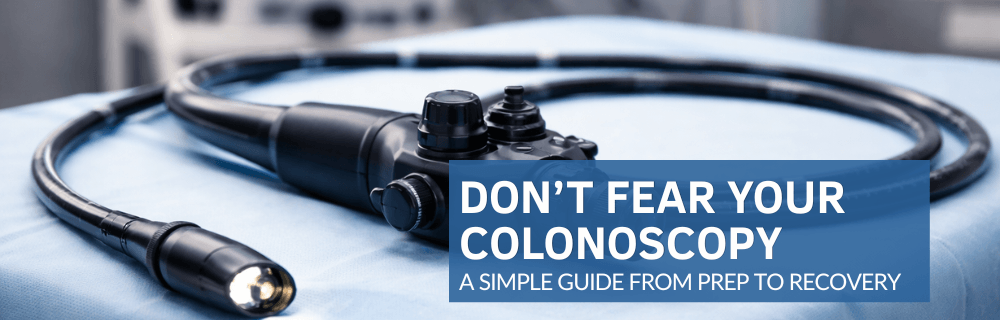
Don’t Fear Your Colonoscopy: A Simple Guide from Prep to Recovery

Don’t Fear Your Colonoscopy: A Simple Guide from Prep to Recovery


For many people, the thought of a colonoscopy can be intimidating. Questions like “Will it hurt?” or “Do I really need this procedure?” often create anxiety.

The truth is, a colonoscopy is a safe, routine procedure that can detect and prevent serious digestive conditions, including colorectal cancer.

At Dr. Deetlefs’s practice, we aim to make the experience as comfortable as possible.

In this guide, we’ll walk you through everything you need to know—from preparation to recovery—so you can approach your colonoscopy with confidence.


What Is a Colonoscopy?


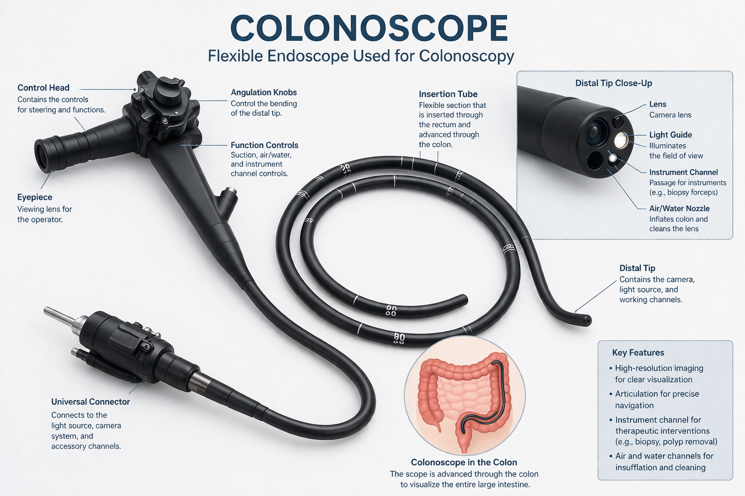
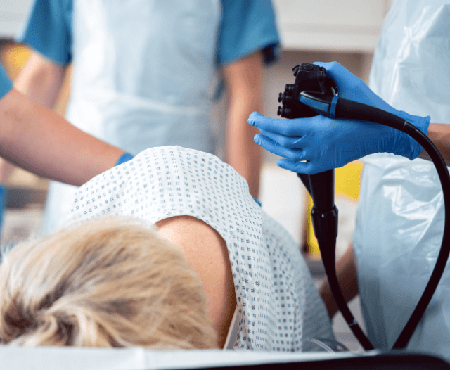
A colonoscopy is a diagnostic procedure that allows a gastroenterologist to examine the inner lining of the colon and rectum. Using a thin, flexible tube with a camera, the doctor can identify:

Dr. Deetlefs emphasizes that early detection via colonoscopy can dramatically increase the likelihood of successful treatment for a range of digestive conditions.


Why Colonoscopies Are Important


Routine colonoscopies are an essential part of preventive digestive healthcare, especially for individuals over the age of 45 or those with a family history of colorectal cancer.

Some key reasons to get a colonoscopy include:

  • Colorectal cancer screening: Detects early-stage cancer and polyps before they become dangerous
  • Investigating digestive symptoms: Unexplained bleeding, chronic diarrhea, constipation, or abdominal pain
  • Monitoring inflammatory bowel disease (IBD): Regular checks help manage conditions like Crohn’s disease or ulcerative colitis

Early detection is crucial. A small polyp can be removed during the procedure, potentially preventing cancer altogether.

colonoscopy_endoscope_procedure


Preparing for Your Colonoscopy


Preparation is often what patients find most challenging, but it is critical for a successful procedure. A clean colon ensures the doctor has a clear view, improving the accuracy of the exam.

Step 1: Dietary Adjustments

A few days before your colonoscopy:

  • Avoid high-fiber foods such as raw vegetables, nuts, and seeds
  • Stick to easily digestible foods like white rice, eggs, and plain chicken

The day before:

  • Switch to clear liquids only, including:
    • Water
    • Clear broths
    • Tea or coffee without milk
    • Clear fruit juices (avoid red or purple)

Avoid alcohol, dairy, and solid foods to ensure your colon is completely clear.

Step 2: Bowel Preparation

Your doctor will provide a laxative solution to drink the day before the procedure. This may cause frequent bowel movements but is necessary to empty your colon. Follow these tips:

  • Stay close to a bathroom
  • Drink plenty of clear fluids to stay hydrated
  • Use moist wipes and barrier creams to reduce discomfort


Step 3: Medication Adjustments

Certain medications may need to be paused, including:

  • Blood thinners
  • Iron supplements
  • Anti-inflammatory drugs

Always follow your doctor’s specific instructions. Never stop medications without consulting your gastroenterologist.


colonoscopy_preparation


What Happens During the Colonoscopy


Most colonoscopies are performed under sedation, so patients feel relaxed and experience minimal discomfort.

Step-by-Step Procedure

  1. Sedation and positioning: You will lie on your side, and a sedative will be administered.
  2. Insertion of the colonoscope: A flexible tube with a camera is gently inserted into the rectum.
  3. Examination of the colon: Air is introduced to expand the colon for better visibility. The camera transmits live images to a monitor.
  4. Polyp removal or biopsy: If polyps or abnormal tissue are found, they can be removed or biopsied immediately.

The entire procedure usually takes 20–45 minutes, depending on the findings. Sedation ensures most patients experience little to no pain.

Is a Colonoscopy Painful?

Thanks to modern sedation techniques, discomfort is minimal. Some patients report:

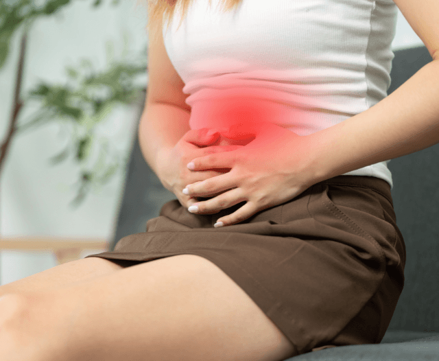
  • Mild cramping
  • A sensation of pressure

These are temporary and generally well-tolerated. Most people are surprised by how quick and smooth the procedure feels.

colonoscopy_polyp_removal


After Your Colonoscopy


Recovery is usually straightforward:

  • You’ll rest until the sedation wears off (typically 30–60 minutes)
  • Mild bloating or cramping may occur due to the air used during the procedure
  • You will need someone to drive you home

Receiving Results

  • Immediate findings may be discussed post-procedure
  • Biopsy results typically take a few days
  • Your doctor will provide guidance on next steps based on your results


Risks and Safety Considerations


Colonoscopy is very safe, but as with any medical procedure, some risks exist:

  • Bleeding, especially after polyp removal
  • Perforation of the colon (rare)
  • Reaction to sedation

Serious complications are uncommon, and the benefits of early detection far outweigh the risks. Discuss any concerns with your gastroenterologist.

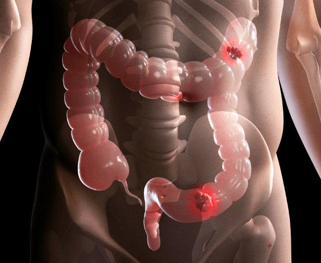
colonoscopy_cancer_screening


When Should You Schedule a Colonoscopy?


You should consider a colonoscopy if you:

  • Are over 45 (routine screening)
  • Have a family history of colorectal cancer
  • Notice persistent digestive symptoms such as bleeding, chronic diarrhea, or unexplained abdominal pain

Even if you feel healthy, routine screening can detect problems before symptoms appear.


Tips for a Comfortable Experience


  1. Follow prep instructions closely: This ensures a smooth procedure.
  2. Ask questions: Knowing what to expect reduces anxiety.
  3. Arrange transportation: Sedation requires a responsible adult to drive you home.
  4. Plan a relaxing day afterward: Most patients feel back to normal by the next day.
  5. Stay hydrated: Drink plenty of clear fluids after your colonoscopy.


Colonoscopy Myths Debunked


“It’s always painful” → Modern sedation makes it almost painless.

  • “Only older people need it” → Family history or symptoms can necessitate earlier screening.
  • “I’ll feel groggy for days” → Most people recover within a few hours.
  • “I don’t need it if I feel fine” → Polyps and early cancers often show no symptoms.


Why Regular Colonoscopy Screening Matters


It’s also important to recognise the long-term value of staying consistent with recommended screening intervals.

A single colonoscopy can provide reassurance for years, especially if no abnormalities are found. For individuals with low risk, follow-up procedures may only be needed every 5 to 10 years, while those with higher risk factors may require more frequent monitoring.

By committing to regular screenings as advised by your gastroenterologist, you are taking a proactive approach to your health—one that can prevent disease progression, reduce the need for more invasive treatments, and ultimately improve your quality of life.

Rather than viewing a colonoscopy as a once-off inconvenience, it should be seen as a strategic investment in your long-term wellbeing.

Cancer_screening_colonoscopy


Take the Next Step Toward Better Digestive Health


If you are experiencing symptoms such as abdominal discomfort, changes in bowel habits, or unexplained bleeding—or if you are due for routine colon cancer screening in South Africa—don’t delay seeking expert care. Early detection is one of the most effective ways to prevent serious digestive conditions and protect your long-term health.

At Dr. Deetlefs’s practice, we are committed to providing personalised, compassionate care in a comfortable and supportive environment. From your initial consultation through to your colonoscopy and follow-up, every step is carefully guided to ensure you feel informed, reassured, and confident in your care.

Our team will walk you through the preparation process, answer any questions you may have, and ensure your procedure is as smooth and stress-free as possible. Whether it’s your first colonoscopy or part of your ongoing health monitoring, you are in experienced hands.

Don’t let uncertainty or fear hold you back.

Take a proactive step today—book your consultation with Dr. Deetlefs and prioritise your digestive health with expert care you can trust.


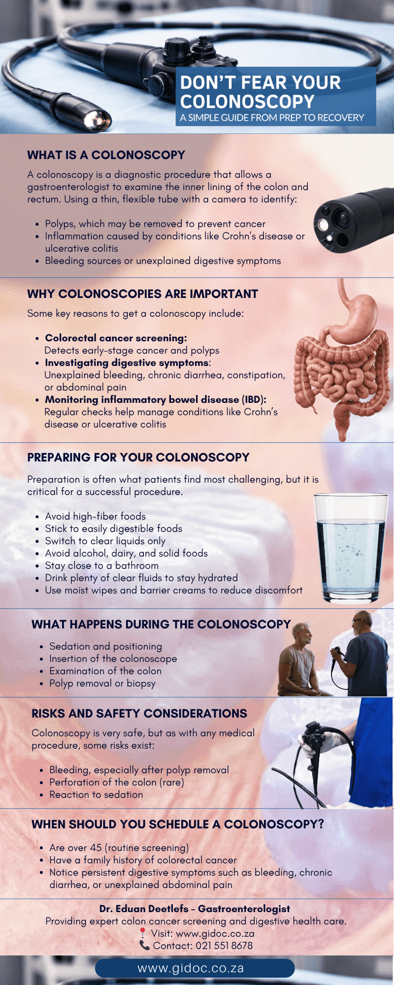
dont_fear_your_colonoscopy_infographic

Conclusion


A colonoscopy is a lifesaving, routine procedure that provides critical insight into your digestive health. Understanding the process—from preparation to recovery—can ease anxiety and empower you to take control of your wellbeing.

At Dr. Deetlefs’s clinic, we prioritize patient comfort, safety, and education. Don’t let fear prevent you from getting the care you need. Early detection can make all the difference.


GIDoc Cape Town

Patient-focused GI treatments and procedures in Cape Town.

Monday-Friday 8AM-4PM.

Connect with Us

© Dr. Eduan Deetlefs, Registered Gastroenterologist, GI Doc Cape Town

Our website information is not intended or implied to be a substitute for professional medical advice, diagnosis or treatment. Please consult a doctor about your specific condition. Only a trained physician can determine an accurate diagnosis and proper treatment.
What Is Endoscopic Dilatation? A Treatment for GI Strictures Explained

What Is Endoscopic Dilatation? A Treatment for GI Strictures Explained

Introduction


Gastrointestinal (GI) strictures—narrowings in the digestive tract—can cause pain, difficulty swallowing, bloating, or bowel obstruction. While these symptoms can severely affect daily life, modern medicine offers effective solutions. One of the most common and minimally invasive treatments is endoscopic dilatation.

At Dr. Deetlefs’s practice, patients with strictures due to conditions like Crohn’s disease, ulcerative colitis, or scar tissue from surgery can benefit from this procedure.

In this article, we explain everything you need to know about endoscopic dilatation, including what it is, how it works, preparation, recovery, and potential risks. 

What Are Gastrointestinal Strictures?

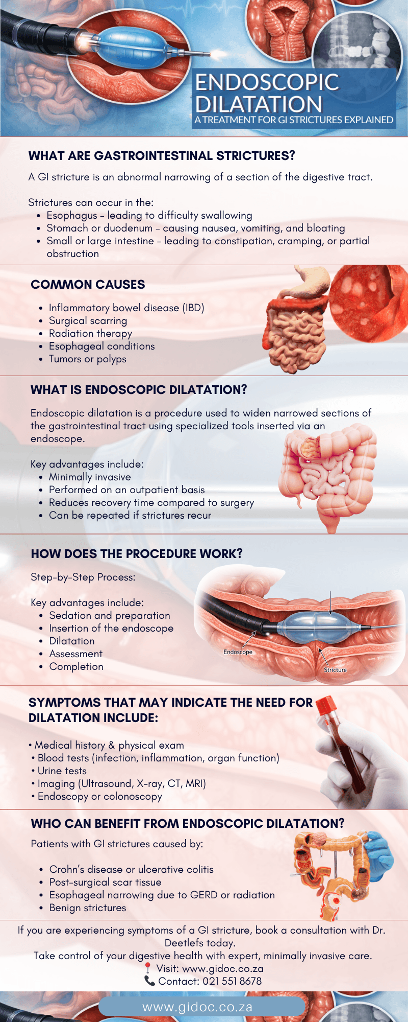
A GI stricture is an abnormal narrowing of a section of the digestive tract. Strictures can occur in the:

  • Esophagus – leading to difficulty swallowing (dysphagia)
  • Stomach or duodenum – causing nausea, vomiting, and bloating
  • Small or large intestine – leading to constipation, cramping, or partial obstruction

Common Causes

  1. Inflammatory bowel disease (IBD)
    • Crohn’s disease is a leading cause of strictures in the small and large intestines
    • Chronic inflammation leads to scar tissue formation
  2. Surgical scarring
    • Post-operative adhesions may narrow sections of the gut
  3. Radiation therapy
    • Can cause tissue scarring in patients treated for abdominal cancers
  4. Esophageal conditions
    • GERD, eosinophilic esophagitis, or acid reflux can lead to esophageal strictures
  5. Tumors or polyps
    • Less commonly, strictures are caused by abnormal growths

What Is Endoscopic Dilatation?


Endoscopic dilatation is a procedure used to widen narrowed sections of the gastrointestinal tract using specialized tools inserted via an endoscope.

The goal is to relieve obstruction, improve swallowing, or restore normal bowel function without the need for major surgery.

Key advantages include:

  • Minimally invasive
  • Performed on an outpatient basis
  • Reduces recovery time compared to surgery
  • Can be repeated if strictures recur

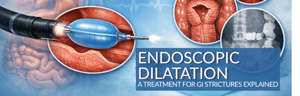
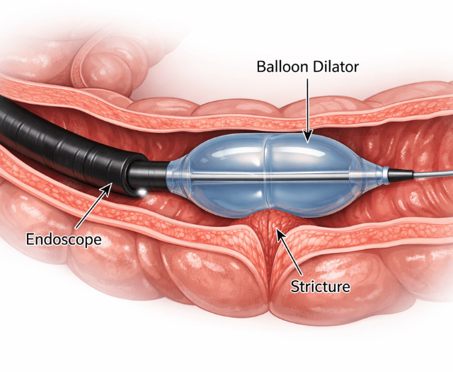
endoscopic_dilation_procedure

How Does the Procedure Work?


Endoscopic dilatation uses either balloon dilators or bougie dilators to gently expand the narrowed area.

Step-by-Step Process

  1. Sedation and preparation
    • The patient receives sedation or anesthesia
    • Positioning depends on the stricture location
  2. Insertion of the endoscope
    • A flexible tube with a camera allows the gastroenterologist to visualize the stricture
    • This ensures precise placement of the dilator
  3. Dilatation
    • Balloon dilator: Inflated gradually at the site of the narrowing to stretch the tissue
    • Bougie dilator: A tapered instrument gently passed through the stricture
  4. Assessment
    • The stricture is reassessed after dilation
    • Multiple passes may be necessary for severe narrowing
  5. Completion
    • The endoscope is removed
    • The patient is monitored briefly before discharge

Endoscopic dilatation is generally safe and effective, with most patients experiencing immediate relief of symptoms.

Who Can Benefit from Endoscopic Dilatation?


Patients with GI strictures caused by:

  • Crohn’s disease or ulcerative colitis
  • Post-surgical scar tissue
  • Esophageal narrowing due to GERD or radiation
  • Benign strictures

Symptoms that may indicate the need for dilatation include:

  • Difficulty swallowing or sensation of food sticking
  • Abdominal pain, bloating, or constipation
  • Vomiting or nausea
  • Weight loss due to poor intake

symptoms_endoscopic_dilation


Preparation for Endoscopic Dilatation


Preparation varies depending on the stricture location:

Esophageal Strictures

  • Fasting: Typically 6–8 hours before the procedure
  • Medication review: Blood thinners or other medications may need temporary adjustment
  • Sedation plan: Discuss anesthesia options with your gastroenterologist

Intestinal Strictures

  • Bowel prep: Cleansing solution may be required for colon or small intestine strictures
  • Dietary instructions: Clear liquids prior to the procedure
  • Medication review: Especially important for patients with IBD

Preparation is critical for safety, visibility, and optimal results.


Recovery After Endoscopic Dilatation


Most patients can return home the same day. Recovery depends on the stricture location and severity.

Immediate Recovery

  • Observation for 1–2 hours post-procedure
  • Mild discomfort, cramping, or bloating may occur
  • Resume clear fluids initially, then gradually progress to regular diet

Short-Term Recovery Tips

  • Eat soft foods for the first few days
  • Avoid hard, crunchy, or sticky foods that may irritate the treated area
  • Stay hydrated

Follow-Up

  • Gastroenterologist may schedule a follow-up endoscopy
  • Multiple dilatations may be necessary if strictures recur
  • Continuous monitoring for patients with IBD is recommended

endoscopic_dilation_recovery


Risks and Complications


Endoscopic dilatation is minimally invasive, but potential risks include:

The procedure is considered safe when performed by an experienced gastroenterologist. Discuss all concerns with your doctor before treatment.


Advantages Over Surgery


Endoscopic dilatation offers multiple benefits compared to surgical intervention:

  1. Minimally invasive – no large abdominal incisions
  2. Short recovery time – most patients resume normal activities within days
  3. Lower risk of complications – less bleeding and infection risk
  4. Repeatable – can be performed multiple times if strictures recur

Surgery may be reserved for severe or complicated strictures that do not respond to endoscopic treatment.


Success Rates and Effectiveness


  • Success rates vary depending on stricture length, location, and underlying cause
  • For short, simple strictures, relief is immediate and long-lasting
  • In patients with Crohn’s disease, multiple sessions may be required due to chronic inflammation

Studies indicate that endoscopic dilatation is highly effective in improving quality of life and preventing obstruction without the need for major surgery (ACG Guidelines).


When to Seek Immediate Medical Attention


After the procedure, contact your gastroenterologist if you experience:

  • Severe abdominal pain
  • Vomiting
  • Fever or chills
  • Significant bleeding
  • Difficulty swallowing or passing stools

Early attention ensures prompt management of any complications.

endoscopic_dilation_complication


Long-Term Management After Endoscopic Dilatation


While endoscopic dilatation provides immediate relief from GI strictures, long-term management is crucial to prevent recurrence.

Patients with conditions like Crohn’s disease or ulcerative colitis should work closely with their gastroenterologist to monitor inflammation, maintain remission, and adjust medications as needed.

Nutritional support is also essential—patients may need a diet rich in easily digestible foods, adequate hydration, and supplementation if malabsorption is present.

Additionally, regular follow-up endoscopies or imaging studies may be recommended to detect early signs of stricture recurrence and address them promptly.

Incorporating lifestyle measures such as stress management, avoiding smoking, and following a tailored anti-inflammatory diet can further improve outcomes and support long-term digestive health.

endoscopic_dilation_infographic


Conclusion


Endoscopic dilatation is a safe, effective, and minimally invasive treatment for gastrointestinal strictures. By widening narrowed areas of the digestive tract, it restores normal function, alleviates symptoms, and can prevent the need for more invasive surgery.

At Dr. Deetlefs’s clinic, we specialize in diagnosing strictures, performing endoscopic dilatation, and managing long-term digestive health.

If you experience difficulty swallowing, persistent bloating, or intestinal obstruction symptoms, don’t wait—early evaluation can improve outcomes and quality of life.

Contact Dr. Deetlefs

If you are experiencing symptoms of a GI stricture, book a consultation with Dr. Deetlefs today.
Take control of your digestive health with expert, minimally invasive care.


GIDoc Cape Town

Patient-focused GI treatments and procedures in Cape Town.

Monday-Friday 8AM-4PM.

Connect with Us

© Dr. Eduan Deetlefs, Registered Gastroenterologist, GI Doc Cape Town

Our website information is not intended or implied to be a substitute for professional medical advice, diagnosis or treatment. Please consult a doctor about your specific condition. Only a trained physician can determine an accurate diagnosis and proper treatment.
Polypectomy: Colon Polyp Removal to Prevent Cancer

Polypectomy: Colon Polyp Removal to Prevent Cancer

Colorectal cancer remains one of the most preventable cancers when detected early. The key lies in identifying and removing precancerous growths—known as polyps—before they transform into malignancy. The procedure used to remove these growths is called a polypectomy, and it is most commonly performed during a colonoscopy.

For many patients, hearing the word “polyp” creates anxiety. However, the majority of colon polyps are benign. The purpose of a polypectomy is preventive: removing abnormal tissue early dramatically reduces the risk of progression to colorectal cancer.

This article explains what polyps are, why they matter, how a polypectomy is performed, what to expect during recovery, and why this procedure plays a critical role in preventive gastroenterology.


What Are Colon Polyps?

Colon polyps are abnormal growths that develop on the inner lining of the colon or rectum. They vary in size, shape, and histological type.

Common Types of Polyps:

  • Adenomatous polyps (adenomas) – These are precancerous and carry the highest malignant potential.
  • Sessile serrated lesions (SSLs) – Also precancerous, often flatter and more subtle.
  • Hyperplastic polyps – Typically benign, especially when small and located in the rectum.
  • Inflammatory polyps – Often seen in inflammatory bowel disease.

Most colorectal cancers arise from adenomatous polyps through what is known as the adenoma-carcinoma sequence, a process that may take 7–10 years. This long window allows for early detection and removal before cancer develops.

polypectomy_procedure


Why Is Polypectomy Important?

A polypectomy is not merely a treatment—it is a cancer prevention strategy.

Large clinical trials have demonstrated that colonoscopy with polypectomy significantly reduces both colorectal cancer incidence and mortality. By removing polyps at an early stage, the progression to invasive cancer is interrupted.

For patients undergoing screening colonoscopy—especially from age 45 onward or earlier in high-risk individuals—polypectomy is often performed during the same session if polyps are detected.


When Is a Polypectomy Performed?

Polypectomy is most commonly performed during a colonoscopy, a procedure that examines the lining of the colon using a flexible camera.

Indications include:

  • Screening colonoscopy
  • Surveillance after previous polyps
  • Investigation of rectal bleeding
  • Evaluation of anemia
  • Unexplained changes in bowel habits
  • Positive stool-based screening tests

If a polyp is identified during colonoscopy, removal is usually performed immediately, provided it is safe to do so.

polypectomy_cancer_screening_follow_up


Preparing for the Procedure

Proper bowel preparation is critical for safe and effective polypectomy.

  • Patients are typically instructed to:
  • Follow a clear liquid diet the day before the procedure
  • Take a prescribed bowel-cleansing solution
  • Avoid certain medications (such as blood thinners) as advised
  • Arrange transportation due to sedation

A clean colon allows for optimal visualization and reduces the risk of missed lesions.


How Is a Polypectomy Performed?

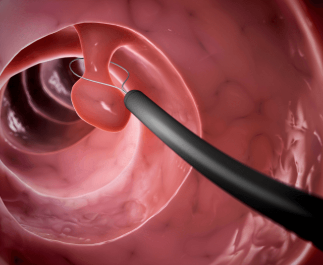
Polypectomy is performed through the colonoscope using specialized instruments. The technique depends on the size, shape, and characteristics of the polyp.

  1. Cold Snare Polypectomy
  • Used for small polyps (typically <10 mm).
  • A thin wire loop (snare) is positioned around the polyp.
  • The polyp is mechanically cut off without electrocautery.

This method minimizes bleeding risk for small lesions.

Cold snaring is now preferred for many small polyps due to its safety profile.

  1. Hot Snare Polypectomy

Used for larger polyps.

  • A snare encircles the polyp.
  • Electrocautery current is applied to cut and cauterize simultaneously.
  • This reduces bleeding risk.

Hot snare techniques are effective for pedunculated (stalked) polyps.

  1. Endoscopic Mucosal Resection (EMR)

For larger, flat, or sessile polyps.

  • Fluid is injected beneath the lesion to lift it from deeper layers.
  • The polyp is then resected using a snare.
  • This technique allows removal of larger lesions without surgery.

EMR is a more advanced technique but avoids the need for open or laparoscopic surgery in many cases.

  1. Piecemeal Resection

Very large polyps may need to be removed in sections. While effective, these cases require close follow-up to ensure complete removal.


polypectomy_polyp_removal


What Happens to the Polyp After Removal?

All removed polyps are sent for histopathological analysis.

The pathology report determines:

  • Polyp type
  • Presence of dysplasia (precancerous change)
  • Margins of resection
  • Need for follow-up surveillance

This information guides future colonoscopy intervals.

Is Polypectomy Painful? 


Patients are sedated during colonoscopy and typically do not feel pain during polypectomy.

After the procedure, mild bloating or cramping may occur due to air introduced during the colonoscopy. This usually resolves within hours.

Most patients resume normal activities the following day.

polyp_removal_polypectomy


Risks and Complications


Polypectomy is generally safe, but potential complications include:

  1. Bleeding
  • Most common complication
  • May occur immediately or up to 7–14 days later
  • Often managed endoscopically
  1. Perforation
  • Rare (less than 1%)
  • Higher risk with large polyps
  • May require surgical management
  1. Post-Polypectomy Syndrome
  • Caused by thermal injury to the colon wall
  • Presents with abdominal pain and fever
  • Usually treated conservatively

Careful technique and experienced endoscopic management significantly reduce these risks.

Recovery After Polypectomy


Post-procedure instructions typically include:

  • Avoid heavy lifting for a few days
  • Resume diet gradually
  • Monitor for bleeding

Seek medical attention if severe pain or persistent bleeding occurs

Minor spotting after bowel movements can occur, especially after removal of larger polyps.

Patients are advised to contact their gastroenterologist if they experience:

  • Heavy rectal bleeding
  • Severe abdominal pain
  • Fever
  • Dizziness or weakness

Surveillance and Follow-Up


Follow-up colonoscopy intervals depend on the number, size, and type of polyps removed.

Examples:

  • 1–2 small adenomas: Repeat colonoscopy in 5–10 years
  • 3–10 adenomas or large lesions: Repeat in 3 years
  • Advanced adenoma: Closer surveillance

Adhering to surveillance guidelines is essential for ongoing cancer prevention. 

polypectomy_procedure_polyp_removal


When Is Surgery Needed Instead?

While most polyps can be removed endoscopically, surgery may be required if:

  • The polyp is too large or invasive
  • Cancer is suspected beyond superficial layers
  • Complete endoscopic removal is not possible

Fortunately, advances in endoscopic techniques have significantly reduced the need for surgical resection in many cases.


Polypectomy and Cancer Prevention


Colorectal cancer often develops silently, without symptoms in early stages. By the time symptoms such as bleeding or weight loss occur, the disease may already be advanced.

Polypectomy interrupts the progression from benign polyp to invasive carcinoma.

This is why routine screening colonoscopy—with removal of detected polyps—remains one of the most powerful preventive interventions in modern medicine.

Who Should Have a Colonoscopy?


General recommendations include:

  • Average-risk individuals starting at age 45

Earlier screening if there is:

  • Family history of colorectal cancer
  • Personal history of polyps
  • Inflammatory bowel disease
  • Genetic syndromes

Early detection saves lives.

polypectomy_polyp_diagnosis


The Role of the Specialist Gastroenterologist


Successful polypectomy requires:

  • Careful lesion detection
  • Appropriate technique selection
  • Complete resection
  • Complication management
  • Structured follow-up planning

An experienced gastroenterologist ensures that polyps are removed safely and thoroughly, minimizing risk and maximizing preventive benefit.

Final Thoughts


A polypectomy may sound intimidating, but it is one of the most effective tools in preventing colorectal cancer. The procedure is typically performed during colonoscopy, is minimally invasive, and has a strong safety profile.

Removing polyps before they become cancerous represents proactive, preventive healthcare at its best.

If you are due for colorectal screening or have been advised to undergo surveillance after previous polyps, consulting a gastroenterologist can provide clarity, reassurance, and potentially life-saving intervention.

Early detection. Complete removal. Ongoing surveillance.
That is the power of polypectomy.

polypectomy_procedure_infographic


Contact Dr. Deetlefs

If you are due for a screening colonoscopy, have previously had colon polyps, or are experiencing concerning bowel symptoms, early evaluation can make all the difference.

Dr. Deetlefs provides comprehensive colorectal cancer screening, expert colonoscopy with polypectomy, and structured follow-up care tailored to your individual risk profile.

Taking a proactive approach today could prevent serious disease tomorrow.

Contact Dr. Deetlefs’ practice to schedule a consultation and take an important step toward protecting your long-term digestive health.


GIDoc Cape Town

Patient-focused GI treatments and procedures in Cape Town.

Monday-Friday 8AM-4PM.

Connect with Us

© Dr. Eduan Deetlefs, Registered Gastroenterologist, GI Doc Cape Town

Our website information is not intended or implied to be a substitute for professional medical advice, diagnosis or treatment. Please consult a doctor about your specific condition. Only a trained physician can determine an accurate diagnosis and proper treatment.