021 202 0626
What is a Polypectomy?

What is a Polypectomy?

A polypectomy is a procedure performed by doctors on patients who have polyps on the inside of their colon, or large intestine.

The polyps are surgically removed in a procedure that is generally considered relatively non-invasive

If you’re a little confused at this point, do not fear.

We’re going to have a look at exactly what polyps are, why they may need to be removed, recovering from a polypectomy and other information that will leave you clued up.

Let’s have a look at what you need to know about a polypectomy.


What are Polyps?


First off, let’s dive into what polyps are. They are, after all, the focus of a polypectomy.

In short, a polyp is an abnormal collection of tissue which grows out of tissue and into hollow spaces in the body.

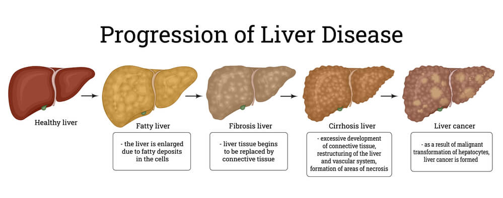
Some types of polyps can change into cancer over time, usually a space of several years, but not all polyps become cancerous. The probability of this depends on the type of polyp.

At times, polyps found through a colonoscopy are likely precancerous. At this stage, they are called adenomas and, once removed, present no further risk to the patient.

Most are benign, however, it is not possible to rule out cancer without first examining the polyp.

It is for this reason that doctors recommend removing polyps when found so that they can be tested for cancer. 

How Common are Polyps?


Polyps are more common than you may think.

Using the United States as an example, between 20-30 percent of Americans have colon polyps.

Uterine polyps are also a problem which increase in probability of occurrence as a woman ages. Resultantly, they are highly prevalent in menopausal women.

Due to their prevalence in urbanized society, it’s imperative to have a screening colonoscopy done at least every 10 years beginning at the age of 50.

The benefits are two-fold: prevent cancer by removing the polyps while they are in the precancerous stage, and assess whether a patient is at risk of developing colon cancer through a lab test of a removed polyp.

If you are thinking that extra tissue such as a polyp may not be a problem if it is benign, think again.

A polyp can grow to a large size.

Such is their growth potential that it can alter blood flow, put pressure on organs, and cause a range of connected side-effects resulting in a multitude of symptoms.


dr-deetlefs-gastroenterology-treatment-capetown


What to Expect in a Polypectomy Procedure


Before doctors can remove a polyp or polyps, they must first ascertain whether there is any present. For this, a procedure called a colonoscopy is used.

During a colonoscopy, a colonoscope is inserted into the patient. A colonoscope is a long, thin and flexible tube which is equipped with a camera and a light at the end.

This allows the doctor to explore the segments of your colon in an attempt to find anything unusual, such as a polyp.

During a colonoscopy, if the doctor discovers polyps, they will usually perform a polypectomy at the same time.

Several methods of undertaking a polypectomy are available to the doctor and this depends in large part on the type of polyps they find.

Polyps can be small, large, sessile, or pedunculated. Sessile polyps don’t have a stalk and lie flat against the colon tissue whereas a pedunculated polyp grows on a stalk and can be thought of as a mushroom in terms of its shape.

Three common polypectomy procedures are as follows:

  • Cold Forceps Polypectomy:
    Forceps are used to pry and pull the polyp loose. The doctor will also use a wire to remove the part of the polyp that extends into the tissue. This technique is most commonly used with small polyps.
  • Hot Forceps Polypectomy:
    This technique is similar to cold forceps removal in that forceps are used to pry and pull the polyp loose. In addition, though, the doctor uses a procedure called electrocautery to burn away any remaining polyp tissue and prevent bleeding.
  • Snare Polypectomy:
    This is the most popular technique used when the polyp is larger than 1cm. A snare is a loop that can grab and remove the polyp. It can be either hot or cold and can be used in conjunction with electrocautery to burn away any remaining polyp tissue.

In moderately rare cases, some polyps can be considered technically challenging or can be associated with an increased risk of complications.

The location and configuration of the polyp, as well as a large polyp size, can result in this scenario.

In these cases, endoscopic mucosal resection (EMR) or endoscopic submucosal dissection (ESD) techniques can be used.

In EMR, a fluid injection made of saline is used to lift the polyp from the underlying tissue and then removed piece by piece.

In ESD, the fluid is injected deep into the lesion and the polyp is removed in one piece.

If this technique is also not possible, such as can be the case with larger polyps, bowel surgery may be needed. This is the last resort, however.


Is Anaesthetic Needed Before a Polypectomy?

Generally, the doctor will give the patient a sedative which is usually administered through an intravenous needle. This helps the patient to feel calm and relaxed.

Anaesthetic is not needed although some patients prefer it.

The procedure itself, while not painful, can be uncomfortable which is why a sedative can help the patient have a more relaxed experience.

In total, the procedure lasts between 30 and 60 minutes and is referred to as an outpatient procedure. This means patients can return home on the same day as the procedure.

In the cases where surgery is required, patients will remain in the hospital for around three days depending on whether complications arise.


Recovery from a Polypectomy


General recovery normally takes a patient around two weeks at a maximum.

In terms of discomfort, patients may feel some pain following the procedure, especially immediately after. The doctor will prescribe medication to manage this pain.

It’s quite normal to experience some bleeding following the procedure but one’s doctor should be kept updated in case there is a complication such as heavy bleeding that stops and starts again.

Your doctor will make sure you are clear on what to do following your procedure with post-care instructions.

They may ask you to avoid certain dietary intake that may irritate your digestive system.

The recommended dietary restriction is normally around 2-3 days.

Some of the foods and liquids the doctor may ask a patient to avoid include coffee, fizzy drinks, alcohol and spicy food.

Apart from the above, your doctor will schedule a follow-up colonoscopy to ensure that the polypectomy was a successful procedure.

 What is a Polypectomy


Summary 

A polypectomy is a routine procedure that can eliminate polyps and prevent the onset of colon cancer as a result of cancerous polyps.

Patients should weigh up the benefits and risks but overall this is a procedure that is relatively non-invasive with a large degree of utility.

For most polypectomy recipients, the procedure is a minor inconvenience that can offer peace of mind with regards to general health.

This becomes especially important when patients are 50 years and older.

If you would like to book an appointment with Dr. Deetlefs  or would simply like more information on a particular GI topic, don’t hesitate to use our online booking form or call us at 021 551 8678.

DISCLAIMER: PLEASE READ CAREFULLY

The information on this website is to provide general guidance. In no way does any of the information provided reflect definitive medical advice and self-diagnoses should not be made based on information obtained online. It is important to consult a Gastroenterologist or medical doctor regarding ANY and ALL symptoms or signs including, but not limited to: abdominal pain, haemorrhoids or anal / rectal bleeding as it may a sign of a serious illness or condition. A thorough consultation and examination should ALWAYS be performed for an accurate diagnosis and treatment plan. Be sure to call a physician or call our office today and schedule a consultation.

GIDoc Cape Town

Patient-focused GI treatments and procedures in Cape Town.

Monday-Friday 8AM-4PM.

Connect with Us

© Dr. Eduan Deetlefs, Registered Gastroenterologist, GI Doc Cape Town

Our website information is not intended or implied to be a substitute for professional medical advice, diagnosis or treatment. Please consult a doctor about your specific condition. Only a trained physician can determine an accurate diagnosis and proper treatment.

Constipation Causes and Treatment in Cape Town

Constipation Causes and Treatment in Cape Town

Have you ever gone for days without feeling a need to go to the toilet?

While some would think that’s normal, most of the time it’s not.

Although there is no generally accepted number of times a person should have bowel movements, there will be times when you’ll notice that you’re visiting the toilet less often than usual.

When you do go to the toilet, it takes all of your strength to pass hard, dry stools.

These are signs of constipation or the condition of having infrequent bowel movements or difficulty in passing stools.

Let’s talk about constipation, what it is, what causes it, and how it can be treated, as well as when you should take it seriously.


constipated_bowel_vs_normal_bowel

How the Bowel Works

To understand why constipation happens, let us first see the normal process of passing stools from the digestive system.

Food travels to the digestive system and is pushed through the intestines which usually takes between 24 to 72 hours.

From the small intestines, where most of the absorption of nutrients takes place, stool consisting of ingested food, bile, and digestive juices is pushed to the large intestines (colon).

The large intestine (colon) is split into four (4) sections: (1) ascending, (2) transverse, (3) descending, and, (4) sigmoid colon. which connects to the rectum and anus.

Food stays within a section for a while, just enough for the digestive tract to absorb fluids and nutrients or process and expel waste.

One of the large intestine’s functions is to absorb most of the fluid that the stool contains to transform it from liquid to solid. The longer this process takes, the more reabsorption occurs, resulting in an increasingly solid stool.

A final bout of reabsorption occurs once the stool reaches the sigmoid colon, before entering the rectum.

The rectal walls will then be distended, signaling the internal anal sphincter to relax. This is the point where the body decides whether to physically expel or hold the stool.

Expelling or retaining the stool in the rectum is controlled by the pelvic floor muscles particularly the puborectalis and external anal sphincter.

The puborectalis forms a single-like formation around the rectum, called the anorectal angle. When you voluntarily relax your external anal sphincter, the stool is finally expelled.

The whole process is coordinated to the brain and is communicated through the sensory nerve pathways that communicate signals such as pain or fullness.

These nerves can also tell the brain if the rectum is filled with gas or stool, which is why you can consciously decide whether to hold or expel it.


Why Constipation Occurs

When you are constipated, a desire to visit the toilet is not enough to signal your body into action, making the whole process of expelling waste more tedious.

This problem usually arises due to two reasons:

  1. The stool’s excessively slow movement through the colon, causing over-absorption of liquid that makes the stool too dry and too hard.
  2. Tightening of the pelvic floor muscles due to pelvic floor dysfunction, aging, or childbirth.

Both problems make the anorectal angle more acute, making it too difficult to expel waste.

 large_intestine_constipation_causes


What is Chronic Constipation?

Going a day without a bowel movement is not necessarily a cause for alarm.

Generally, constipation becomes chronic when you defecate less than three times per week for several months.

Chronic constipation interferes with people’s ability to go about their daily tasks and can cause excessive straining and extreme discomfort during bowel movements.

Signs and symptoms of chronic constipation include:

  1. Bowel movement that occurs less than three (3) times per week
  2. Consistent passing of lumpy hard stools
  3. Continuous straining to have bowel movements
  4. Feeling of blockage in the rectum
  5. Feeling as though you cannot empty the rectum
  6. Needing help to empty the rectum by pressing the abdomen or using the finger to remove stool from the rectum.

If you have experienced two or more of these symptoms for the last two or three months, constipation may be considered chronic. It’s best to book an appointment with your Gastroenterologist right away.

 causes-of-constipation

Causes of Constipation

Chronic constipation has many possible causes:

1. Blockages in the colon or rectum

A blockage in the colon or rectum can slow down or completely stop stool movement.

Blockages may be caused either by anal fissures or tiny tears in the skin around the anus, bowel obstruction in the intestines, colon or rectal cancer and other abdominal cancers that presses on the colon, bowel stricture or narrowing of the colon, rectocele or a rectum bulge through the back wall of the vagina.


2. Damaged nerves around the colon and rectum

Since nerves have a vital role in the transmission of stool, it’s natural for neurological problems to cause issues with bowel movements.

Autonomic neuropathy or damaged nerves, multiple sclerosis, Parkinson’s disease, spinal cord injury, and stroke are some of the conditions that bring about chronic constipation.

3. Pelvic muscle problems

Weakened pelvic muscles that are unable to relax or contract correctly can cause chronic constipation.

4. Underlying conditions affecting hormones

Underlying conditions affecting hormones such as diabetes, overactive thyroid (hyperthyroidism), underactive thyroid gland (hypothyroidism), and pregnancy may cause a prolonged bowel movement.

Aging, pregnancy, dehydration, low-fiber diet, sedentary lifestyle, medications (e.g., sedatives, pain relievers, anti-depressants, blood pressure control), and some mental health conditions (e.g., eating disorder, depression) can increase one’s risk of developing chronic constipation.


Complications of Chronic Constipation

If chronic constipation remains untreated, the patient may develop the following complications:

1. Hemorrhoids – These are swollen veins in the anus that are caused by continuous straining to have a bowel movement. Hemorrhoid pain can be sudden and severe. The patient might feel or see a lump around the anus.

2. Anal fissure – This is a torn skin in your anus that is caused by a large or hard stool.

3. Fecal impaction – This is the accumulation of hardened stool that gets stuck in the intestines and cannot be expelled.

This is a very serious condition and needs medical attention right away. Fecal impaction won’t go away on its own. You need to have it removed for you to get better.

4. Rectal prolapse – This occurs when the intestine starts protruding from the anus due to continuous straining. Surgery is needed to repair the prolapse.


Diagnosis and Treatment for Constipation

in Cape Town

Treating constipation starts with diagnosing and trying to find the cause. Several tests can be made including the following:

  • Blood tests – Your doctor will watch for systemic conditions such as hypothyroidism or high calcium levels.
  • X-ray – This will show if your intestines are blocked and if there is stool present throughout the colon.
  • Sigmoidoscopy – Examination of the rectum and lower, or sigmoid, colon
  • Colonoscopy – Examination of the rectum and entire colon
  • Colonic transit study – You may be asked to swallow a capsule with a wireless recording device or radiopaque marker. The progress of the capsule through your colon will be recorded for 24 to 48 hours and will be shown on X-rays.

Constipation Causes and Treatment in Cape Town


Once the underlying cause is determined, your Gastroenterologist will recommend an appropriate treatment plan. Treatment may include:

1. Lifestyle changes: The doctor may recommend simple lifestyle changes such as stress management, daily exercise, and quitting smoking.

2. Laxatives: These are drugs that trigger muscle contractions in the small intestines to help advance bowel movement.

3. Fiber supplements: If the patient is found lacking in fiber, fiber supplements may be helpful to aid in digestion and defecation.

4. Biofeedback: Assessment and measurement of muscle tension in the rectum to teach physical techniques that improve pelvic muscle performance so the patient can pass stool more easily.

5. Pain management: This focuses on relieving the pain associated with chronic constipation.

6. Surgery: Some cases may need surgery, especially if there are structural issues in the small intestines such as rectocele.

If you’re experiencing signs of chronic constipation, seek medical advice right away to avoid complications.

Schedule an appointment with Dr. Deetlefs in Cape Town, South Africa to get prompt medical assistance for chronic constipation and other issues with your digestive health.


DISCLAIMER: PLEASE READ CAREFULLY

The information on this website is to provide general guidance. In no way does any of the information provided reflect definitive medical advice and self-diagnoses should not be made based on information obtained online. It is important to consult a Gastroenterologist or medical doctor regarding ANY and ALL symptoms or signs including, but not limited to: abdominal pain, haemorrhoids or anal / rectal bleeding as it may a sign of a serious illness or condition. A thorough consultation and examination should ALWAYS be performed for an accurate diagnosis and treatment plan. Be sure to call a physician or call our office today and schedule a consultation.

GIDoc Cape Town

Patient-focused GI treatments and procedures in Cape Town.

Monday-Friday 8AM-4PM.

Connect with Us

© Dr. Eduan Deetlefs, Registered Gastroenterologist, GI Doc Cape Town

Our website information is not intended or implied to be a substitute for professional medical advice, diagnosis or treatment. Please consult a doctor about your specific condition. Only a trained physician can determine an accurate diagnosis and proper treatment.

What is (PEG) Placement of Feeding Tubes

What is (PEG) Placement of Feeding Tubes

EG stands for percutaneous endoscopic gastrostomy.

It is a procedure in which a flexible feeding tube is placed through the abdominal wall and into the stomach.

PEG allows nutrition, fluids and/or medications to be put directly into the stomach, bypassing the mouth and oesophagus.

When Does One Require a PEG?

If you have trouble swallowing or can’t eat or drink enough through your mouth, you may just need a feeding tube. You may get one through your nose or mouth for a few days or weeks while you recover from an illness.

But if you have long-term or serious reasons why you’re unable to eat, such as dementia or terminal cancer, you may need to undergo a fairly simple surgery called a PEG.

Your surgeon generally cuts through the skin of your belly and inserts the tube right into your stomach to deliver a liquid food mixture or a formula.

How is the PEG Performed?

Your doctor will use a lighted flexible tube called an endoscope to guide the creation of a small opening through the skin of the upper abdomen and directly into the stomach.

This procedure allows the doctor to place and secure a feeding tube into the stomach.

Patients generally receive an intravenous sedative and local anaesthesia, and an antibiotic is given by vein before the procedure.

Patients can usually go home the day of the procedure or the next day.


3 Different PEG Placements

There are, however, three different methods in which a PEG can be placed.

The method used by your doctor will depend on a variety of factors, the most common technique being the aforementioned endoscopic placement.

1.    Endoscopic

Even within this method, there are variations in methodology. The most common is referred to as the “Push and Pull Technique” and is the method that will be described below.

The doctor needs to be assisted by another operator (apart from the normal support staff).

Before the PEG tube placement, an antibiotic is given to the patient.

Once this has been done, the doctor will make sure there are no obstructions, such as oesophagal obstruction or an ulcer at the site where the PEG tube would be placed, that may get in the way of the PEG tube placement. This is achieved by performing an upper endoscopy.

With the endoscope in the stomach, the second operator will find an appropriate point on the abdomen by looking for transillumination (light from the endoscope inside the stomach) through the abdominal wall.

The position is confirmed when the second operator presses on the chosen area with one finger, and a clear indentation of the gastric wall is seen by the doctor with the endoscope.

This technique that combines the second operator pressing with a finger and the doctor looking for the point of the press with the endoscope increases the safety of PEG tube insertion.

Once an appropriate site on the abdomen is selected, the abdominal wall is cleaned with an iodine-based solution, and the second operator dons sterile gloves.

A local anaesthetic is injected at the site. A small skin incision may then be made at the site (approximately 1 cm in length).

The medical team will then insert the PEG tube going through several technical steps.

2.    Radiological

Radiologically-inserted gastrostomy tubes are placed using fluoroscopic-guidance. Radiologic insertion of a gastrostomy tube requires that the stomach be distended (swollen) with air.

This is often done with a nasogastric tube (one that goes to the stomach via the nose) though alternative techniques may also be used.

Once the stomach is distended, an appropriate site is chosen using fluoroscopy (typically the mid-body of the stomach). A 2-cm square around the site is designated, and a local anaesthetic is injected at the four corners.

The doctor then performs a gastropexy to prevent the stomach from moving up into the chest.

Following this, the tube is placed after some more anaesthetic, an incision, and a couple more technical steps.

3.    Surgical

Surgical gastrostomy can be carried out either laparoscopically (minimally invasive) or using an open approach.

Laparoscopic gastrostomy tube placement is performed similarly to radiologic gastrostomy tube placement.

After the laparoscope is inserted, the gastrostomy site is chosen and grasped. T-fasteners are then used to affix the stomach to the abdominal wall, and the gastrostomy tube is placed.

Open insertion of a gastrostomy tube involves the creation of a midline incision. The stomach is located, and a gastric incision is made.

percutaneous_ endoscopic_gastrostomy

The gastrostomy tube is then inserted, and the gastric and midline incisions are closed.

The Day Before Your Procedure

Instructions for eating and drinking before PEG Surgery

  • Do not eat anything after midnight the night before your surgery. This includes hard candy and gum.
  • Between midnight and up until 2 hours before your scheduled arrival time, you may drink a total of 300ml of water.
  • Starting 2 hours before your scheduled arrival time, do not eat or drink anything. This includes water.

How Should I Care for the PEG Tube?

  • Always wash hands before handling your PEG tube.
  • Clean the site with soap and water daily. DO NOT use hydrogen peroxide or any special cleansers. You may use a q-tip or gauze to swab gently around the site. Rinse well and pat dry. This may be done in the shower.
  • Apply a clean dressing to the site. This should be changed daily or as needed. This dressing should be placed over the external bumper.
  • When changing the dressing, look for redness on your skin, drainage on the old dressing, or leakage at the site of insertion. If found, report to your doctor.
  • Tape the tube to your skin to prevent tugging on the skin leading to skin breakdown. Paper tape works well and is gentle on your skin.
  • Turn the bumper (or sometimes called a bolster, the piece of soft plastic that sits on the skin to prevent the tube from slipping back into the stomach) and tube halfway at least twice a day to prevent skin breakdown. Also gently push and pull the tube in and out – 1/2 cm each day.
  • Flush tube with 30 ml of water at least once a day.

 tube_feeding_after_peg

How are Feedings Given? Can I Still Eat and Drink?

Specialized liquid nutrition, as well as fluids, are given through the PEG tube.

If the PEG tube is placed because of swallowing difficulty (e.g. after a stroke), there will still be restrictions on what you can take in orally.

Although a few PEG patients may continue to eat or drink after the procedure, this is a very important issue to discuss with your doctor as instructions will vary based on your specific case.

Complications from PEG Placement

While not common, complications can occur with the PEG placement.

Possible complications include pain at the PEG site, leakage of stomach contents around the tube site, and dislodgement or malfunction of the tube.

Possible complications include infection of the PEG site, aspiration (inhalation of gastric contents into the lungs), bleeding and perforation (an unwanted hole in the bowel wall).

Your doctor can describe symptoms that could indicate a possible complication.

How long do these tubes last? How are they removed?

PEG tubes can last for months or years. This, naturally, varies case-by-case.

However, because they can break down or become clogged over extended periods, they might need to be replaced.

Your doctor can easily remove or replace a tube without sedatives or anaesthesia, although your doctor might opt to use sedation and endoscopy in some cases.

Your doctor will remove the tube using firm traction and will either insert a new tube or let the opening close if no replacement is needed.

PEG sites close quickly once the tube is removed, so accidental dislodgement requires immediate attention.

PEG vs. PEJ

When preparing for a PEG tube placement, you may hear the term “PEJ”. Let’s clear up any possible confusion around the terms ‘PEG’ and ‘PEJ’.

A PEG tube is a feeding tube that is placed in your stomach.

If the tube can’t be placed into your stomach, you may have a PEJ tube placed instead. A PEJ tube is placed in your jejunum, which is the second part of your small intestine.

The tube is placed during an endoscopy (a procedure that lets your doctor see inside your stomach and small intestine).

A PEJ tube placement is, therefore, a similar procedure to a PEG tube placement with the only difference being that the tube gets inserted into the jejunum instead of the stomach.

If you need long-term nutrition support, your doctor may convert your PEG into a low-profile gastrostomy button into your stomach.

Your doctor may convert your PEJ into a low-profile jejunostomy button into your small intestine. With both of these buttons, you will attach the feeding adapter to get nutrition.

PEG - Placement of Feeding Tubes

Summary

A PEG tube placement is a fairly straightforward procedure.

It may sound complicated to a first-time reader but it’s standard practice to help patients who have feeding complications and shouldn’t be feared.

If you would like to book an appointment with a gastrointestinal (GI) specialist or would simply like more information on a particular GI topic, don’t hesitate to use our online booking form or call us at 021 551 8678.


DISCLAIMER: PLEASE READ CAREFULLY

The information on this website is to provide general guidance. In no way does any of the information provided reflect definitive medical advice and self-diagnoses should not be made based on information obtained online. It is important to consult a Gastroenterologist or medical doctor regarding ANY and ALL symptoms or signs including, but not limited to: abdominal pain, haemorrhoids or anal / rectal bleeding as it may a sign of a serious illness or condition. A thorough consultation and examination should ALWAYS be performed for an accurate diagnosis and treatment plan. Be sure to call a physician or call our office today and schedule a consultation.

GIDoc Cape Town

Patient-focused GI treatments and procedures in Cape Town.

Monday-Friday 8AM-4PM.

Connect with Us

© Dr. Eduan Deetlefs, Registered Gastroenterologist, GI Doc Cape Town

Our website information is not intended or implied to be a substitute for professional medical advice, diagnosis or treatment. Please consult a doctor about your specific condition. Only a trained physician can determine an accurate diagnosis and proper treatment.

Gastrointestinal Bleeding Lesions: Causes, Diagnosis and Treatment

Gastrointestinal Bleeding Lesions: Causes, Diagnosis and Treatment

Pronounced “lee-sion” with an emphasis on the “lee,” a lesion can be almost any damage or abnormal change involving any tissue or organ due to physical trauma or disease.

The word “lesion” comes from the Latin noun “laesio” meaning “an attack or injury” which is related in Latin to the verb “laedere” = “to hurt, strike or wound.”

Because the definition of a lesion is so broad, the varieties of lesions are virtually endless.

Lesions can occur anywhere in the body and are generally classified by their patterns, their sizes, their locations, or their causes.

Here we look at some of the ways in which lesions vary from one another and then we will take a closer look at gastrointestinal (GI) lesions and their treatment process.

1. Cancerous Lesions

Lesions can be categorized according to whether or not they are caused by cancer. A benign lesion is non-cancerous whereas a malignant lesion is cancerous.

For example, a biopsy of a skin lesion may show that the lesion is benign or malignant.

It’s also possible to tell if the lesion is in a state of evolving into a malignant lesion. In this case, it would be called a pre-malignant lesion.

2. Variations in Lesion Patterns

Lesions can be defined according to the patterns they form.

The particular pattern which a lesion forms can also form the basis of it’s naming convention.

A bullseye, or target, lesion, for example, is one that looks like the bullseye on a target.

An example of its identification is through that of an X-ray of the duodenum where the bullseye lesion can represent a tumour with an ulcer in the centre.

Another distinct, patterned lesion is that of the coin lesion. This lesion is a round shadow resembling a coin on a chest X-ray It, too, is usually due to a tumour.


3. Lesions Named After People

In typical human fashion, we like to name many things after ourselves.

Lesions are not exempt from this. Lesions can be named after the person who first described them.

For instance, a Ghon lesion (or Ghon focus) is the scar-like “signature” in the lungs of adults left by tuberculosis in childhood.

4. Variations in Lesion Sizes

Lesions also vary by name and categorization based on size.

A gross lesion is one that can be seen with the naked eye.

A microscopic or histologic lesion requires the magnification provided by a microscope to be seen.

Lesions on the molecular level have also been classified. The basis of sickle cell disease, for example, is a molecular lesion as it is only detectable on the DNA level.


5. Location-Based Lesions

Location is another basis for naming lesions. In neurology, a central lesion involves the brain or spinal cord, i.e., the central nervous system.

This is in contrast to a peripheral lesion which involves the nerves away from the spinal cord and does not involve the central nervous system.

6. Many Different Names for Many Different Lesions

There is a virtually endless assortment of lesions in medicine:

Primary lesions, secondary lesions, impaction lesions, indiscriminate lesions, irritative lesions, etc.

Many are named after people including the Armanni-Ebstein lesion, a Bankart lesion, a Blumenthal lesion, and so on. 

gastrointestinal_lesions

Gastrointestinal Lesions

As we’ve seen, a lesion can be almost any damage or abnormal change involving any tissue or organ due to physical trauma or disease.

A gastrointestinal lesion can often result in bleeding. A symptom of a lesion, or general disorder in your digestive tract, blood may appear in your stool or vomit although it isn’t always visible.

The level of bleeding can range from mild to quite severe so it is important to know how to read the signs so that you may get help as soon as possible.

Your doctor may use sophisticated imaging technology to locate the cause of bleeding followed by varying treatment depending on the gastrointestinal bleeding.


Causes of Gastrointestinal Bleeding Lesions

If bleeding occurs in your oesophagus, stomach, or the initial part of the small intestine (duodenum), it’s considered upper GI bleeding.

Bleeding in the lower small intestine, large intestine, rectum, or anus is called lower GI bleeding.


Causes of Upper GI Bleeding

Peptic ulcers are a common cause of GI bleeding. These ulcers are open lesions that develop in the lining of your stomach or duodenum.

An infection from H. pylori bacteria usually causes peptic ulcers.

Another cause of upper GI bleeding can be the result of enlarged veins in your oesophagus tearing. This is a condition called oesophageal varices.

Tears in the walls of your oesophagus can also cause GI bleeding.

A quite serious cause of upper GI bleeding is that of Dieulaofoy’s Lesion which occurs in the stomach wall. While rare, it can be life-threatening and must be ruled out when GI bleeding occurs.


Causes of Lower GI Bleeding

Colitis, inflammation of the colon, is the most common cause of lower GI bleeding. Colitis has multiple causes, including infection, food poisoning, and Crohn’s disease.

Haemorrhoids are another common cause of GI bleeding. A haemorrhoid is an enlarged vein in your rectum or anus. These enlarged veins can rupture and bleed.

symptoms_of_bleeding_lesions

Symptoms of GI Bleeding

There are a few things that you can look for if you suspect that you might have GI or rectal bleeding.

Your stool might become darker and sticky, like tar, if bleeding comes from the stomach or upper GI tract.

You may pass blood from your rectum during bowel movements, which could cause you to see some blood in your toilet or on your toilet tissue.

This blood is usually bright red. Vomiting blood is another sign that there’s bleeding somewhere in your GI tract.

If you experience any of these symptoms call your doctor immediately.

GI bleeding could signal a life-threatening condition and, as such, immediate medical treatment is essential.

Also, seek treatment immediately if you experience any of the following symptoms:

  • shortness of breath
  • paleness
  • weakness
  • dizziness

These symptoms may also signal severe bleeding.


Symptoms of Shock

If your bleeding starts abruptly and progresses rapidly, you could go into shock. Signs and symptoms of shock include:

  • Drop in blood pressure
  • Not urinating or urinating infrequently, in small amounts
  • Rapid pulse
  • Unconsciousness


When to See a Doctor?

If you have symptoms of shock, you or someone else should call your emergency medical number.

If you’re vomiting blood, see blood in your stools or have black, tarry stools, seek immediate medical care. For other indications of GI bleeding, make an appointment with your doctor.


Request an Appointment with Dr Deetlefs


treatment_of_bleeding_lesions


Diagnosis of GI Bleeding

Diagnosis of the underlying cause of your GI bleeding will usually start with your doctor asking about your symptoms and medical history.

Your doctor may also request a stool sample to check for the presence of blood along with other tests to check for signs of anaemia.

Upper GI bleeding is most commonly diagnosed after your doctor performs an endoscopic examination.

Endoscopy is a procedure that involves the use of a small camera located atop a long, flexible endoscopic tube your doctor places down your throat.

The scope is then passed through your upper GI tract.

The camera allows your doctor to see inside your GI tract and potentially locate the source of your bleeding.

Because endoscopy is limited to the upper GI tract, your doctor may perform an enteroscopy. This procedure is performed if the cause of your bleeding isn’t found during an endoscopy.

An enteroscopic exam is similar to an endoscopy, except there’s usually a balloon attached to the camera-tipped tube. When inflated, this balloon allows your doctor to open up the intestine and see inside.

To determine the cause of lower GI bleeding, your doctor may perform a colonoscopy. During this test, your doctor will insert a small, flexible tube into your rectum.

A camera is attached to the tube so your doctor can view the entire length of your colon.

Air moves through the tube to provide a better view. Your doctor may take a biopsy for additional testing.

 lesion_prevention_treatment

Treatment for GI Bleeding Lesions

An endoscopy can be useful, not only in diagnosing GI bleeding but also for treating it.

The treatment is called haemostasis of bleeding lesions and the idea is to stop the bleeding.

The use of special scopes with cameras and laser attachments, along with medications can be used to stop the bleeding.

In addition, your doctor can use tools alongside scopes to apply clips to the bleeding vessels to stop the bleeding.

If haemorrhoids are the cause of your bleeding, over-the-counter (OTC) treatments might work for you.

If you find that OTC remedies don’t work, your doctor might use a heat treatment to shrink your haemorrhoids.

If infection is the cause then the doctor might prescribe antibiotics which are usually enough to treat infections.


How to Prevent a Lesion?

You cannot always predict and therefore prevent lesions.

There are some lesions which you can reduce the probability of occurrence of. Gastrointestinal lesions, for example, may be less likely to occur if you follow a healthy lifestyle.

By “healthy lifestyle” I mean the age-old yet perfectly-sound advice of the following:

  1. Follow a regular exercise routine.
  2. Limit your use of non-steroidal anti-inflammatory drugs.
  3. Eat according to a healthy meal plan with lots of fruit and vegetables.
  4. Avoid smoking and drinking alcohol.
  5. Establish and stick to a consistent sleep routine.
  6. Reduce stress where possible through practices such as meditation.

 gastrointestinal-lesions


Treating GI Bleeding Lesion


Gi Doc Capetown is a practice founded to provide medical solutions in the area of gastroenterology.

Over the years, Dr Eduan Deetlefs has earned the reputation of a trusted health expert providing consultative, diagnostic, and therapeutic endoscopic and related services to patients in Cape Town and beyond. 

DISCLAIMER: PLEASE READ CAREFULLY

The information on this website is to provide general guidance. In no way does any of the information provided reflect definitive medical advice and self-diagnoses should not be made based on information obtained online. It is important to consult a Gastroenterologist or medical doctor regarding ANY and ALL symptoms or signs including, but not limited to: abdominal pain, haemorrhoids or anal / rectal bleeding as it may a sign of a serious illness or condition. A thorough consultation and examination should ALWAYS be performed for an accurate diagnosis and treatment plan. Be sure to call a physician or call our office today and schedule a consultation.

GIDoc Cape Town

Patient-focused GI treatments and procedures in Cape Town.

Monday-Friday 8AM-4PM.

Connect with Us

© Dr. Eduan Deetlefs, Registered Gastroenterologist, GI Doc Cape Town

Our website information is not intended or implied to be a substitute for professional medical advice, diagnosis or treatment. Please consult a doctor about your specific condition. Only a trained physician can determine an accurate diagnosis and proper treatment.

Fatty Liver Disease: Causes, Symptoms, and Treatments

Fatty Liver Disease: Causes, Symptoms, and Treatments

After a long week’s work, all you want to do on a Friday night is to eat out, drink, and have fun with your colleagues or friends.

The night gets long, you go home tired, and while resting, your liver works harder.

The liver is the body’s largest internal organ which is about the size of a football. The liver is the body’s blood purifier.  Without it, our bodies will be filled with toxins that will make us sick.

Its primary functions are to:

  • filter and get rid of the toxins and other blood impurities;
  • produce and excrete bile that helps with digestion by breaking down fat in the small intestine;
  • make blood plasma proteins;
  • convert excess glucose into glycogen for storage that can be later turned back into glucose for energy as needed;
  • manage blood clotting; and
  • metabolize fats, proteins, and carbohydrates.

Because of these functions, the liver is one of the organs that are most subject to abuse and damage. One of the most common liver diseases is the fatty liver disease (FLD) or hepatic steatosis.

What is Fatty Liver Disease?

FLD is a condition where excess fat builds up in the liver. It happens when the liver cannot metabolize fat, resulting in the fat not being excreted out of the body.

There are two types of FLD: non-alcoholic fatty liver disease (NAFLD) and alcoholic fatty liver disease (AFLD). By the names of it, NAFLD is acquired by those who drink little to no alcohol and the latter is from consuming too much alcohol over time.

Fatty liver is actually the earliest stage of liver disease.

Having a fatty liver is not life-threatening, but neglecting this condition can lead to other more serious conditions such as liver cirrhosis and even cancer.


What are the Causes and Risk Factors of FLD?


The excess or build-up of fat in the liver can be caused by various habits and medical conditions. The causes can be:

  • Alcohol Abuse
    When metabolizing alcohol in the liver, toxic metabolites are produced. This commonly happens to chronic alcoholics.
  • Metabolism-Related Disease
    FLD can be caused by existing disorders such as abetalipoproteinemia or the disorder when proper absorption of fat and fat-soluble vitamins from food is obstructed.Another example is glycogen storage disease when the synthesis or metabolism of glycogen is affected by enzyme deficiencies.Because heh body is an interconnected system of organs, problems with metabolism can affect the liver and cause FLD and other liver diseases.
  • Nutrition-Related Factors
    Obesity or excess accumulation of fat in the body can also cause FLD. Other factors include high blood sugar, insulin resistance, and high levels of triglycerides in the blood.Triglycerides are also known as lipids or a type of fat. When you eat, the calories that your body does not immediately need are converted to triglycerides and stored in fat cells. These are later released for your body’s energy in between meals.Rapid weight loss and malnutrition can also cause FLD.
  • Drugs and Toxins Intake
    Fatty liver disease can sometimes be a side effect of medications and drug intake, as well as exposure to toxins.

While chronic alcohol intake is one of the major causes of FLD, some people develop FLD even when they do not have the mentioned habits and medical conditions.
Some factors increase the risk of having a fatty liver including the following:

  • obesity
  • chronic viral hepatitis
  • genetics
  • old age
  • removed gallbladder
  • polycystic ovary syndrome
  • sleep apnea
  • type II Diabetes
  • hypothyroidism
  • hypopituitarism

How Serious is a Fatty Liver?

If fat is detected in the liver biopsy but the liver is not swollen and has no tissue damage, the diagnosis may be non-alcoholic fatty liver disease (NAFLD).

This is a simple fatty liver and nothing serious. But you need to treat it before it develops into serious liver diseases.

Non-alcoholic steatohepatitis or NASH is one of the serious liver diseases.
It is diagnosed if you have fat in the liver, and the organ is swollen and damaged.
When liver damage from NASH is left untreated, it can lead to much more serious liver diseases like cancer.

Permanent scarring (fibrosis) to the liver that causes it to harden is cirrhosis or liver cancer.

In a 2010 study of NAFLD in Western Cape, South Africa, 48% of the 233 screened patients had NAFLD, of whom 36% had NASH and 17% had advanced liver fibrosis.


What are the Symptoms of FLD?

FLD usually gives no symptoms. People often learn about their fatty liver because of medical exams for other reasons or during their annual physical and medical tests.

The liver is situated in the upper right abdomen, below the lungs, and on top of the stomach. This is the part that throbs, hurts, or feels tired that may indicate damage to the liver.

In cases of fatty liver disease that has developed into a serious condition like non-alcoholic steatohepatitis (NASH), the following symptoms may be experienced:

  • Swollen belly
  • Enlarged bloodvessels underneath the skin that appear to be spiderlike
  • Larger-than-normal breasts in men
  • Red palms
  • Skin and eyesthat appear yellowish.


How is FLD Diagnosed?

You may experience no symptoms that will directly relate to fatty liver disease so it is best to have yourself checked annually or whenever you deem necessary.

Detection of a probable FLD can be through a blood sample or an imaging test.

If there is something unusual in the blood test results or the liver appears slightly enlarged in an imaging test, your doctor may recommend the following procedures to diagnose FLD:

  • Lifestyle and Health History
    Your doctor may ask about your alcohol drinking habits, eating habits, and if you have relatives who had liver diseases. Your doctor may also ask about your other medical conditions that may have caused FLD.
  • Imaging Test
    Fat in the liver appears bright in an ultrasound. Other imaging tests such as CT scans or magnetic resonance imaging can also show fat in the liver.
  • Physical Exam
    Your doctor may inspect your body and look for symptoms such as swollen belly or jaundice, a condition that causes a yellow pigmentation in the skin and the sclera (white part in the eyes), among others.


  • Serology or blood test.
    High levels of specific enzymes indicate a problem in the liver.
    Serology is also used to eliminate possibilities of other diseases such as hepatitis.
  • Liver biopsy.
    Your doctor may recommend this if a serious condition of fatty liver disease is suspected.
    In a liver biopsy, a sample of liver tissue is collected and examined.

It is also best to talk to your doctor about other symptoms you may have been experiencing like fatigue, loss of appetite, etc.

You can also ask your doctor about clarifications and the next steps if you will be diagnosed with FLD.


Fatty Liver Disease_cape_town


How is Fatty Liver Disease Treated?

A simple case of fatty liver disease can be treated by correcting habits and making lifestyle changes to control fat accumulation in the liver.

A proper and balanced diet is a good start. You also need to decrease your calorie intake, cut back on your sugar, and exercise regularly.

Avoid alcohol or drink in moderation whether you have alcoholic or non-alcoholic fatty liver disease.

Always check your medicines or prescriptions from other conditions before mixing them with alcohol. Mixing drugs and alcohol can cause damage to the liver.

If you drink heavily or think you are suffering from alcoholism, you can confidentially disclose this to your doctor.

This way, your medication and treatment can address both the fatty liver disease and alcoholism and other issues that may hamper your recovery and affect your overall health.

Contact Dr. Deetlefs today for an appointment in Cape Town.

DISCLAIMER: PLEASE READ CAREFULLY

The information on this website is to provide general guidance. In no way does any of the information provided reflect definitive medical advice and self-diagnoses should not be made based on information obtained online. It is important to consult a Gastroenterologist or medical doctor regarding ANY and ALL symptoms or signs including, but not limited to: abdominal pain, haemorrhoids or anal / rectal bleeding as it may a sign of a serious illness or condition. A thorough consultation and examination should ALWAYS be performed for an accurate diagnosis and treatment plan. Be sure to call a physician or call our office today and schedule a consultation.

GIDoc Cape Town

Patient-focused GI treatments and procedures in Cape Town.

Monday-Friday 8AM-4PM.

Connect with Us

© Dr. Eduan Deetlefs, Registered Gastroenterologist, GI Doc Cape Town

Our website information is not intended or implied to be a substitute for professional medical advice, diagnosis or treatment. Please consult a doctor about your specific condition. Only a trained physician can determine an accurate diagnosis and proper treatment.