021 202 0626
What is EMR Polypectomy?

What is EMR Polypectomy?

You may have heard of the word “polyp” before.

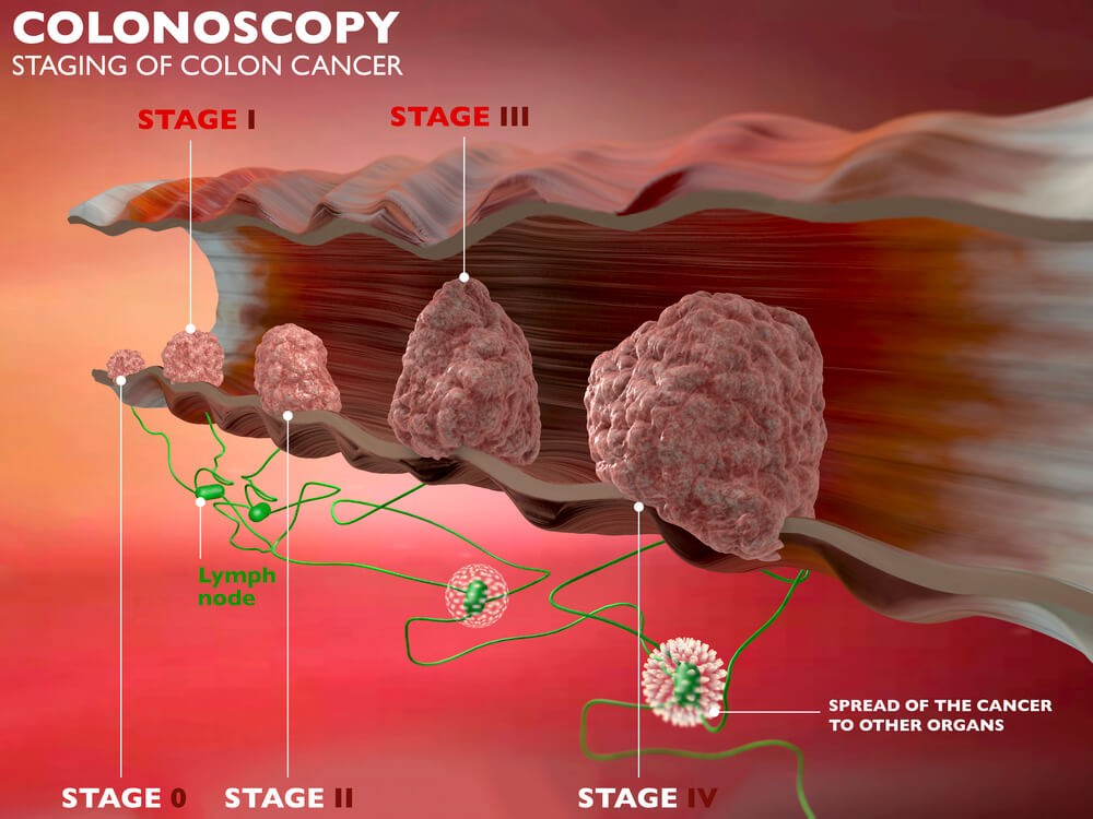
Polyps are abnormal tissue growths that most commonly develop in the colon. They look like small bumps and may even resemble mushroom stalks.

While most polyps are benign (non-cancerous), because it’s an abnormal tissue growth, there’s a high chance it can become cancerous and cause you tons of problems later on.

That’s why polyps need to be examined and removed as soon as possible.

That’s what the EMR polypectomy is for.


EMR Polypectomy

Endoscopic Mucosal Resection, or EMR, is a procedure that involves the removal of polyps or tumors in the digestive tract which may be precancerous or at early stage cancer.

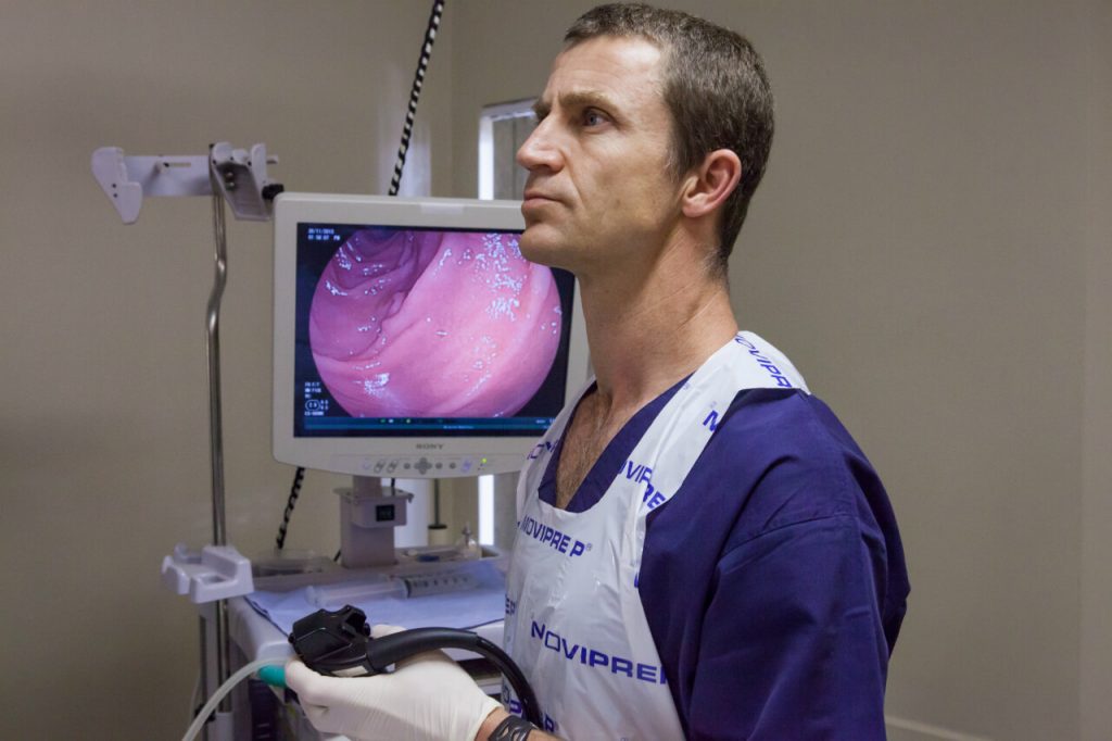
The instrument used in EMR polypectomy is an endoscope – a flexible narrow tube with a light and a video camera at the end that allows the gastroenterologist to see the inside of your body with elaborate details.

The endoscope is passed down to the patient’s throat to reach the site of the polyp either in the patient’s esophagus, stomach, or duodenum (the upper part of the small intestine).

The endoscope may also be guided up through the anus if the site of abnormality is found in the colon.
EMR is a curative procedure, but it may also be used for in-depth diagnostic and staging purposes as EMR is also useful in biopsy tissue sampling.

EMR also helps in determining whether cancer cells have already invaded tissues beneath the digestive tract lining.

EMR is also used to treat Barrett esophagus – a condition in which the cells that make up your esophagus begin to look like the cells that make up your intestines as cells were constantly exposed to acid due to years of gastroesophageal reflux (GERD).  


What is the Difference between EMR and ESD?

EMR is most effective for tumors that have not yet reached deeper layers of the gastrointestinal wall and are no larger than two (2) centimeters.

If the tumor has already reached deeper layers, your gastroenterologist may suggest an endoscopic submucosal dissection or ESD, a procedure used to remove deep tumors from the GI tract.


What Happens During an EMR Polypectomy?

EMR is a minimally invasive procedure that does not involve an incision.

Because of this, patients usually recover faster and experience less pain compared to open or laparoscopic surgery.

As it is usually done as an outpatient procedure, you will be able to go home the same day unless your doctor recommends otherwise.

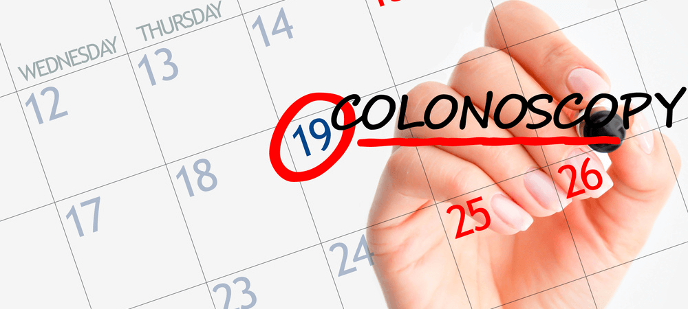
Patients will be asked to arrive at the endoscopy unit three or more hours before the procedure to register and provide your medical history, including all taken medications.


Before the Procedure

Your doctor should give you written instructions on how to prepare for the EMR.

This usually involves fasting and emptying the colon, which is extremely important for your doctor to clearly see what’s going on inside your body.

You will also need to go on a strict clear liquid diet the day before the procedure.

A liquid laxative or an over-the-counter enema kit might also be prescribed to help empty out the colon.

Once you arrive at the clinic, a nurse will ask you some questions and fill up the necessary paperwork. You will then be asked to change into a gown.

Your pulse and blood pressure will be checked, and a small needle will be inserted into the back of your hand.

Your doctor should also explain to you the procedure, the benefits and risks, and answer any questions that you may have.

You will be asked to sign a consent form to confirm your understanding of what the gastroenterologist has explained and to signify your consent to the procedure.


emr_polypectomy

During the Procedure

You will be asked to remove false teeth or glasses before lying down on a trolley on your left or right side, whichever is comfortable for you.

Your doctor will give you sedation to make you feel relaxed and drowsy.
Pain relief will also be administered.

The sedation is not a general anaesthetic. You will still be able to hear the nurses/doctor talking to you. However, the effect of sedation may cause you to not remember the procedure afterward.

You will also be given oxygen to aid in breathing.

The endoscope will be gently inserted through the mouth/anus, wherever the location of the tumor, and images of your GI tract will be observed by the doctor on the screen.

Sometimes, applying a suction to the top of the tumor is necessary to further lift the tumor up away from the other tissue.

This prevents creating further damage to the surrounding tissues during the procedure.

Your doctor will then insert a thin wire rope through the endoscope to cut the tumor at its base and seal the cut at the same time.

The tumor will then be retrieved through suction and pulling through a specialized retrieval tool that will be passed through the endoscope.

A laboratory examination will confirm if the tumor has been completely removed from the GI tract.


After the Procedure

After the procedure, you will be transferred and monitored in a recovery room while the sedative wears off.

Occasionally, the sedative lowers the blood pressure and may also cause breathing problems. This makes it important to closely monitor your pulse and blood pressure before being cleared to go home.

Before you leave the facility, your gastroenterologist will discuss after-care and the next steps you need to take.

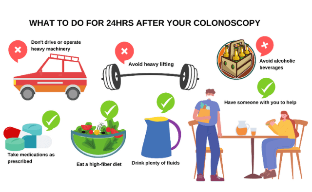
You shouldn’t drive (vehicle insurance will become invalid), operate any machinery, drink alcohol, or sign any legally binding documents within 24 hours after the procedure.

At home, you may drink or eat as normal, but try to eat a high-fiber diet.

Some of the after-effects of EMR include slight bloating and bleeding, sore throat, nausea, and vomiting. These are normal and should subside within 24 hours. If not, call your doctor immediately.


What are the Risks of EMR?

EMR is proven to be a safe and effective technique in the management of GI lesions when performed by an experienced gastroenterologist or endoscopist, given that it is applied only on an appropriate lesion.

Bleeding is the most common complication during EMR that can be detected and corrected even during the procedure.

There’s an extremely slight chance (1 in every 1500 people) for you to suffer a perforation or significant bleeding that requires an operation and/or blood transfusion.

There is also a slight risk of narrowing of the esophagus which may lead to difficulty in swallowing and may require further treatment.

There may also be a slight risk of puncture on the site where the lesion has been removed.

If you develop fever, chills, vomiting, black stool, bright red blood in the stool, chest or abdominal pain, shortness of breath, and/or fainting after undergoing EMR, you must seek medical attention immediately.

With that said, the chances for these complications to happen are very slim. When properly done, the benefits of EMR far outweigh the risks.

                               EMR-Polypectomy-Cape-Town

Follow-Up Appointment

You will likely have a follow-up appointment with your doctor to discuss the results and findings of the EMR.

It is important to ask your doctor if they were able to remove all the abnormal tissues and if there were any cancerous among those tissues.

Three to twelve (3-12) months after the procedure, a follow-up exam may be performed to make sure that all abnormal lesions have been completely removed from the GI tract.

EMR polypectomy is a safe procedure that has saved millions of lives.

In the care of a good gastroenterologist, the procedure should go smoothly without any hitch.

If you need a trusted gastroenterologist to perform your EMR polypectomy in Cape Town, give Dr. Deetleefs a call today.


DISCLAIMER: PLEASE READ CAREFULLY

The information on this website is to provide general guidance. In no way does any of the information provided reflect definitive medical advice and self-diagnoses should not be made based on information obtained online. It is important to consult a Gastroenterologist or medical doctor regarding ANY and ALL symptoms or signs including, but not limited to: abdominal pain, haemorrhoids or anal / rectal bleeding as it may a sign of a serious illness or condition. A thorough consultation and examination should ALWAYS be performed for an accurate diagnosis and treatment plan. Be sure to call a physician or call our office today and schedule a consultation.

GIDoc Cape Town

Patient-focused GI treatments and procedures in Cape Town.

Monday-Friday 8AM-4PM.

Connect with Us

© Dr. Eduan Deetlefs, Registered Gastroenterologist, GI Doc Cape Town

Our website information is not intended or implied to be a substitute for professional medical advice, diagnosis or treatment. Please consult a doctor about your specific condition. Only a trained physician can determine an accurate diagnosis and proper treatment.

Liver Disease: Symptoms and Treatment

Liver Disease: Symptoms and Treatment

The liver is one of the most important organs in our bodies with a key role in digesting food and getting rid of toxic substances.

This is why liver failure is one of the worst conditions you can have.

It compromises your immune system, causes deterioration of brain functions, may lead to kidney failure, and, in many cases, even death.

The good news is a liver disease can be prevented by simple lifestyle changes. Liver disease can also be successfully treated with early detection and proper medical intervention.  

liver-anatomy


Anatomy of the Liver

The liver is known as the second-largest organ in the body. An adult’s liver can weigh approximately 3 pounds and can be as large as a football.

It’s reddish-brown in color and sits just under your rib cage on the right side of your abdomen.

The liver is divided into two sections, the right and left lobes.

The right lobe receives blood primarily from the intestine while the left lobe receives blood coming from the stomach and the spleen.

Both lobes are made up of 8 segments which are comprised of a thousand small lobes called “lobules”.

These lobules connect to a network of small ducts (tubes) that also connect with larger ducts to ultimately form the network of the hepatic duct.

Bile, a clear yellow or orange fluid produced by the liver to help break down fats and digest food, passes through the hepatic duct down to the gallbladder and the duodenum, the first section of the small intestine.


The Primary Functions of the Liver

The liver is a powerhouse that performs over 500 life-sustaining functions.

Its main function is to filter the blood coming from the digestive tract before it passes through the rest of your body.

The body cannot perform its metabolic processes without the liver. Nutrients cannot be absorbed by the body without the liver.

Even medicines cannot be broken down and be used by the body without passing through the liver.

Here is a list of the primary metabolic processes the liver performs:

1. Produces bile, which helps carry away waste products and break down fats to prepare them for further digestion and absorption

2. Produces blood plasma from certain proteins. Blood plasma carries nutrients, hormones, water, salts, enzymes, and proteins to different body parts. Cells also deposit their waste into the plasma

3. Produces cholesterol and special proteins to transport fats through the body

4. Stores and releases glucose as the body needs it

5. Stores and uses iron from processing hemoglobin

6. Changes harmful ammonia to urea to be excreted through urine

7. Filters the blood of medicines and other toxic substances.

8. Regulates blood clotting.

9. Produces immune factors and removes bacteria from the bloodstream to help the body fight infections.

10. Clears blood from bilirubin, a compound that causes the skin and eyes to turn yellowish when it reaches higher-than-average levels.

 Liver-Disease-Symptoms-Treatment-infographic


Liver Disease: Stages, Diagnosis, and Treatment

The liver has amazing abilities to self-repair. In fact, it is the only organ in the body that can grow back after parts of it were removed or damaged.

However, repetitive abuse may lead to liver failure and even death.

Liver disease is caused by a variety of factors – genetics, viruses, or, most commonly, unhealthy lifestyle choices (e.g. alcoholism and obesity).

Over time, these conditions may lead to the scarring of the liver (cirrhosis), which can lead to liver failure.

Detecting liver disease in its early stages increases the chances of reversing the damage, so it’s important to know what symptoms to watch out for.


stages-of-liver-damage


4 Stages of Liver Disease

There are different kinds of liver disease but with any liver disease, the symptoms and liver damage will progress in a similar way. 

STAGE 1: An Inflamed Liver

Having an enlarged or inflamed liver is a sign that your immune system is responding to foreign substances (i.e. toxins) in the body.

There could be excess fats, too many toxins, or a viral infection that is causing your liver to swell up.

A non-alcoholic fatty liver, a fatty liver, liver hepatitis, viral hepatitis, or autoimmune hepatitis, are some of the conditions that may bring about an inflamed liver.

A patient with an inflamed liver reports stomach pain or a burning sensation at the upper right area of the abdomen; however, in some cases of non-alcoholic fatty liver, there will be no symptom of inflammation at all.

See your Gastroenterologist immediately if you have felt any sign of liver damage. Treating your liver at this stage can help reverse liver damage.


STAGE 2: Fibrosis – The Start of Liver Scarring

If the liver is left inflamed, fibrosis will take place. Fibrosis is the process where scarring in the liver starts due to constant inflammation.

The scar tissues will replace healthy liver tissues and will eventually reduce liver function.

The scar tissues will also cause toxins and fat to continually build up and in turn block blood to flow to the organ.

At this stage, you’ll still have a chance to save your liver through medication and lifestyle management.


normal-liver-vs-liver-with-cirrhosis


STAGE 3: Cirrhosis – Most Severe Scarring

Not treating fibrosis will lead to the development of cirrhosis, which is a severe scarring of the liver. 

At this stage, the liver can no longer heal itself and thus may lead to many complications including cancer.

Here are some of the symptoms or complications:

  1. Constant fatigue
  2. Itching
  3. Loss of appetite
  4. Sudden weight loss
  5. Nausea
  6. Bloating of the abdomen
  7. Edema or swelling due to a build-up of fluid in the feet, ankles, or legs
  8. Spider-like blood vessels found on the skin
  9. Jaundice – yellowing of the skin and eyes

The treatment of liver disease done at this stage is to control the progress of scarring, as well as dealing with any complications and symptoms that may arise from cirrhosis.


STAGE 4: Liver Failure

Unfortunately, at this stage, your liver has lost all its ability to function, making it unable to heal.

Your physician will immediately give you medical attention to salvage what is left of your liver; but if this is no longer possible, the only remaining option is to have a liver transplant.


Diagnosis of Liver Disease

If you have been experiencing any of the previously mentioned symptoms, it’s crucial to schedule an appointment with a doctor as soon as possible.

A thorough examination of your health history and physical examination will help find the cause and extent of liver damage.

Your doctor may ask you to undergo several liver function tests through blood extraction.

Other blood tests might also be done to look for specific liver problems or genetic conditions.

Imaging tests such as ultrasound, CT scan, and MRI might also be recommended to show liver damage.

A biopsy (removal of a tissue sample from your liver) might also be conducted to further look for signs of liver disease or liver damage.


human-liver-infographic


How to Care for Your Liver

Lifestyle changes cannot reverse cirrhosis but they can help to delay or stop the progression of liver disease and help prevent further complications.

Here are some of the lifestyle changes that can help you care for your liver while it’s not yet too late:

1. Start Eating Healthy

It all starts and ends in nutrition. Eat a balanced and healthy diet, limit your consumption of processed foods, detoxify your system by staying hydrated, and avoid sugary and salty food.

2. Quit Smoking

Cigarettes contain a host of toxins that can pose a serious toll on the health of your liver as the liver is the primary organ that filters toxins from our blood.

3. Avoid Drinking Alcohol

Alcohol is your liver’s worst enemy. Constant consumption of alcohol above the recommended limit can lead to severe liver damage or failure.

4. Manage Stress

Not many realize that stress can affect one’s overall health. Stress is like toxins not just to our mind but also to our organs. To make your liver healthy again, make sure to avoid getting stressed or find effective stress management techniques.

5. Regularly Exercise

Regular exercising and maintaining a healthy body weight lowers the chance of fat accumulation in the liver.

Our liver is one of the hardest working organs in our body as it works overtime to rid our bodies of toxins.

Maintaining a healthy lifestyle and seeking medical advice as soon as symptoms of liver damage occur are keys to keeping our livers healthy well into our senior years.

DISCLAIMER: PLEASE READ CAREFULLY

The information on this website is to provide general guidance. In no way does any of the information provided reflect definitive medical advice and self-diagnoses should not be made based on information obtained online. It is important to consult a Gastroenterologist or medical doctor regarding ANY and ALL symptoms or signs including, but not limited to: abdominal pain, haemorrhoids or anal / rectal bleeding as it may a sign of a serious illness or condition. A thorough consultation and examination should ALWAYS be performed for an accurate diagnosis and treatment plan. Be sure to call a physician or call our office today and schedule a consultation.

GIDoc Cape Town

Patient-focused GI treatments and procedures in Cape Town.

Monday-Friday 8AM-4PM.

Connect with Us

© Dr. Eduan Deetlefs, Registered Gastroenterologist, GI Doc Cape Town

Our website information is not intended or implied to be a substitute for professional medical advice, diagnosis or treatment. Please consult a doctor about your specific condition. Only a trained physician can determine an accurate diagnosis and proper treatment.

Routine Colonoscopy Screening: Important Information

Routine Colonoscopy Screening: Important Information

Colorectal cancer (CRC) is cancer that occurs in the colon or rectum, where abnormal growths called polyps form.

According to the World Cancer Research Fund, colorectal cancer is the third (3rd) most common histologically diagnosed cancers worldwide.

This ranking is also true in South African cancer cases among women, while it ranks as the

second (2nd) leading cause of cancer deaths among South African men, which, only has as little as 0.1% survival rate.

A routine colonoscopy screening might be just what you need to not join the statistics.


What is a Routine Colonoscopy Screening?

A colonoscopy is a procedure doctors conduct to screen for colorectal cancer and can also help diagnose unexplained changes in bowel habits and abdominal pain.

Even without symptoms, undergoing a routine colonoscopy can help your doctor locate polyps so they can be removed before turning into cancer.

It aids doctors to find colorectal cancer at an early stage when a patient’s survival rate is still at its highest.

As it is a visual exam, colonoscopy screening involves the use of a colonoscope and a proctosigmoidoscope to look at the entire length of the colon and rectum.

These instruments are thin fiber optic devices with a light and video camera that are attached to the tip of a flexible tube to search for polyps.

The devices are passed through the anal orifice into the rectum and further inside into the colon. Mild sedation and pain medication are usually administered in this procedure.

Also, sspecial instruments can be passed through the colonoscope to get a biopsy (sample) or remove any suspicious-looking growths such as polyps if needed.


How Often Do I Need to Undergo Colonoscopy Screening?

Everybody should undergo a complete colonoscopy screening by the time they reach the age of forty-five.

By this age, the overall risk of developing colorectal cancer increases and early detection is vital for effective treatment.

If you are forty-five (45) years old or older, with an average risk of CRC you may have to routinely undergo colonoscopy once every 10 years.

People at increased or high risk of colorectal cancer might need to start colorectal cancer screening before age 45, be screened more often, and/or get specific tests.

As shown in this table, some indicators increase a person’s risk of developing colorectal cancer, and a higher risk requires more frequent colonoscopy screening:


INDICATORS Average Risk of CRC High Risk of CRC
  Colonoscopy requirement   Colonoscopy requirement
Personal history of colorectal cancer or certain types of polyps every 10 yrs. Regular colonoscopy, 1 year after previous surgery
Family history of colorectal cancer every 10 yrs. Before reaching age 45
Personal history of inflammatory bowel disease (ulcerative colitis or Crohn’s disease) every 10 yrs. Colonoscopy at least 8 years after being diagnosed with inflammatory bowel disease. Follow-up colonoscopies should be done every 1 to 3 years, depending on the person’s risk factors for colorectal cancer and the findings on the previous colonoscopy.

Personal history of getting radiation to the abdomen (belly) or pelvic area to treat a prior cancer


every 10 yrs. Colonoscopy at an earlier age (depending on how old they were when they got the radiation); often begins 5 years after the radiation was given or at age 30, may need to be screened more often than normal (such as at least every 3 to 5 years).
With genetic disease every 10 yrs. Colonoscopy as early as teenage years


Colonoscopy Routine Screening vs. Diagnostic Colonoscopy

Screening colonoscopy and diagnostic colonoscopy basically perform the same procedures and only differ in purpose and timing.

While screening colonoscopy is a preventive measure to screen patients for cancerous polyps when they reached a certain age or if they have a family history of colon cancer, diagnostic colonoscopy is more of a curative response.

Diagnostic colonoscopies are done when a patient already exhibits specific symptoms that may suggest colon cancer, such as persistent diarrhea or constipation; blood in the stool; persistent abdominal cramps, gas or pain; weakness or fatigue; unexplained weight loss; and that feeling of not being able to empty completely no matter how many bowel movements.


stages-of-colon-cancer


It is important to note that most colorectal cancer patients experience no symptoms in the early stages of the disease.

When symptoms start to show, cancer cells have most likely grown within the large intestine; therefore, it is important that you get screened.


Preparation: Things to Do to Prepare for a Colonoscopy

For your colonoscopy to be a success, you need to endure the hardship of purging as you need to empty your colon.

You will be prescribed a powerful bowel-clearing laxative resulting in frequent bowel movements or diarrhea both the night before or the morning of the procedure.

Failure to empty your colon may cause the polyps and lesions to be missed, the colonoscopy may take longer (increasing the risk of complications), or the whole process may need to be delayed and rescheduled – which means you will undergo another round of bowel prep (which you wouldn’t wish to).

Your doctor may ask you to refrain from ingesting solid food and only intake water the day before your procedure.

If you have pre-existing conditions like diabetes, heart problems, or high blood pressure, or if you have been on medication or supplements with iron, let your physician know at least a week before the screening.

Also inform your doctor if you have been taking aspirin, warfarin (Coumadin, Jantoven), anticoagulants like dabigatran (Pradaxa) or rivaroxaban (Xarelto). You might be asked to adjust your dosages or stop taking the medications temporarily.


During: What Happens During a Colonoscopy

The whole procedure typically lasts approximately 30-60 minutes and may take longer if the doctor sees abnormalities that need removal.

Many people are given medicines so they are put into a sleepy state where they can still converse but they won’t have any memory of the conversation.

You may still feel mild cramping during the procedure which you can reduce by taking several slow deep breaths. Some people, however, may need stronger anesthesia.

You will be asked to lie on your left side on the examining table as the doctor will insert the colonoscope through the rectum and advance to the other end of the large intestine while the instrument transmits images of the colon lining to the screen.

The scope is bendable so the doctor can move it around the curves of your colon. You may be asked to change position as needed to facilitate the moving of the scope.

Your colon expands as the scope blows air into your colon so the doctor can see more clearly.

When the doctor has finished, he will slowly withdraw the colonoscope while carefully examining the lining of your bowel.

after_treatment_colonoscopy

Recovery: What to Do After a Colonoscopy

It is particularly important that you are accompanied by someone in the next 24 hours after your procedure.

As you have been sedated, it is not recommended that you drive, fly, or do any strenuous activity.

Watch your diet, drink plenty of fluids, and stick to your prescribed medications.

If polyps have been removed in your colon, you might need to take at least 7 days to rest and alter your activities including exercising 

Possible Risks of Colonoscopy

Although rare, some complications may arise after a colonoscopy. Studies estimate the overall risk of complications for routine colonoscopy to be as low as 1.6%.

Perforation (a hole in the intestine), bleeding, post-polypectomy syndrome, reaction to anesthetic and infection are some of the possible complications that may occur.

Despite these, colonoscopy is an overall very safe test, and the benefits of undergoing routine colonoscopy screening far outweigh the discomfort and possible risks that may arise.

We highly recommend that you get screened because as the saying goes, “prevention is better than cure”.

DISCLAIMER: PLEASE READ CAREFULLY

The information on this website is to provide general guidance. In no way does any of the information provided reflect definitive medical advice and self-diagnoses should not be made based on information obtained online. It is important to consult a Gastroenterologist or medical doctor regarding ANY and ALL symptoms or signs including, but not limited to: abdominal pain, haemorrhoids or anal / rectal bleeding as it may a sign of a serious illness or condition. A thorough consultation and examination should ALWAYS be performed for an accurate diagnosis and treatment plan. Be sure to call a physician or call our office today and schedule a consultation.

GIDoc Cape Town

Patient-focused GI treatments and procedures in Cape Town.

Monday-Friday 8AM-4PM.

Connect with Us

© Dr. Eduan Deetlefs, Registered Gastroenterologist, GI Doc Cape Town

Our website information is not intended or implied to be a substitute for professional medical advice, diagnosis or treatment. Please consult a doctor about your specific condition. Only a trained physician can determine an accurate diagnosis and proper treatment.

Haemostasis Treatment in Cape Town

Haemostasis Treatment in Cape Town

Any abnormal internal and external bleeding is a cause of serious concern.
When left untreated, it may cause complications and even death.

If you are suffering from a bleeding disorder, it’s best to undergo a medical examination and get the appropriate treatment right away.

But what is the science behind uncontrolled bleeding? Let’s talk about haemostasis and its important role in regulating bleeding.


What is Haemostasis?

Haemostasis is the first stage of wound healing. It is the medical term for the body’s response to preventing and stopping bleeding when there is an open wound.


haemostasis-blood-coagulation


The whole process involves coagulation, blood changing from liquid to gel, to close the open hole and start repairing the blood vessel.

A healthy person’s bleeding will automatically stop after a short time to prevent extensive blood loss.

It is a vital physiological function that keeps us from losing too much blood volume.

If severe blood and fluid loss occur, the heart will pump insufficient blood carrying oxygen to the body causing tissue and organ damage. This medical emergency is called a hypovolemic shock.


Haemostasis and Bleeding Disorders


Bleeding disorders are a set of disorders that are characterized by defects in haemostasis, inability to perform proper blood clots, which lead to an increased susceptibility to bleeding (“haemorrhagic diathesis”).

These disorders can lead to heavy and prolonged bleeding after experiencing injury, trauma, or menstruation.

Bleeding disorders may be inherited or acquired and are either caused by

(1) platelet disorders (primary haemostasis defect) – either too few or abnormal platelets, or,

(2) coagulation defects (secondary haemostasis defect) – abnormal or low amounts of clotting proteins,

(3) abnormal blood vessels, or, in some cases, a combination of two or all the three.

Haemophilia is perhaps the most familiar inherited bleeding disorder, although it is relatively rare and mostly affects males.

However, statistics show that the most recurring blood disorder is the von Willebrand disease, caused by clotting proteins.

Both male and female are susceptible to this disease and is usually acquired rather than inherited.


Some other symptoms of a bleeding disorder that needs your immediate attention are easy bruising, bleeding gums, heavy bleeding from small wounds or dental work, unexplained nosebleeds, heavy menstrual bleeding, bleeding into joints, and excessive bleeding following surgery.

Make sure to consult with a physician if you exhibit some of these symptoms.
Treatment for bleeding disorders varies, depending on the condition and its severity.

For some bleeding disorders, there are topical products, nasal sprays, and fresh frozen plasma that might need to be administered while in hospital confinement.

For other bleeding disorders, you may be medicated at home with clotting factor concentrates that can be infused prophylactically.


Internal Bleeding

External bleeding caused by cuts, abrasions, or nose bleeds can be easily located and addressed, which is not the case for internal bleeding.

There are internal bleedings that may become visible like

(1) a bruise, when blood from damaged blood vessels leaks into the surrounding skin;

(2) bleeding from the anus that implies bowel injury;

(3) bleeding from the ears or nose that imply a head injury;

(4) coughing up frothy bloodied spit that implies a lung injury might have occurred; and,

(5) blood found in the urine which implies a urinary tract injury.

Internal bleeding is a medical emergency with serious, even fatal, consequences. Prompt medical help is vital, as first aid cannot manage or treat any kind of internal bleeding.


Gastrointestinal Bleeding

If you or someone you know is vomiting blood, has blood found in his/her stools, or have black, tarry stools, we highly recommend seeking immediate medical care as this is likely to be a case of gastrointestinal bleeding.

Gastrointestinal (GI) bleeding is any bleeding in the GI tract and may be found either in the mouth, oesophagus, stomach, small intestines, large intestines, or the anus.

The degree of bleeding might be from mild to severe or life-threatening. 

haemostasis-treatment-cpt


Microscopic bleeding can be detected only by lab tests which can lead to anaemia over time, if not detected.

Perceptible amounts of bleeding that can be seen in the stool or vomit are treated as a medical emergency as it can lead to death.

GI bleeding is commonly caused by a gastric or peptic ulcer, duodenal ulcer, or pancreatic disease, among others.


Role of Endoscopic Haemostasis Therapy in the Management of Gastrointestinal (GI) Bleeding

Acute gastrointestinal bleeding is among the most urgent situations in daily gastroenterological practice. Endoscopy plays a key role in the diagnosis and treatment of GI bleeding cases.

Endoscopic haemostasis is probably the most important technical challenge that must be mastered by gastroenterologists.

It is crucial for both the prevention of bleeding and the management of gastrointestinal bleeding during high-risk endoscopic procedures.

Its primary aim is to control bleeding or reduce the likelihood of further bleeding.

Its role is not limited to primary detection of GI bleeding, but also to establishing the severity of a lesion and providing relevant information regarding its risk of bleeding.

Endoscopic haemostasis involves a combination of techniques to stop gastrointestinal bleeding.

It is very crucial to correctly identify the source of bleeding as approximately 2% of lesions may not be identified at the first endoscopy.

Once the source of bleeding is established, the application technique and haemostasis and the initiation of the pharmacological treatment will be the next step.


Techniques in Endoscopic Haemostasis

New endoscopic techniques, accessories, and compounds are now available to make this minimally invasive treatment suitable for a vast array of digestive lesion types.

The main techniques used for endoscopic treatment of digestive bleeding are

(1) endoscopic ligation,

(2) injection techniques (i.e. adrenaline or sclerosing substances injection),

(3) thermal techniques, and

(4) mechanical techniques (use of metal clips, sewing equipment or equipment for mechanical suture).


Endoscopic Ligation

Endoscopic ligation is a procedure that uses elastic bands to treat veins and varices that were enlarged in the oesophagus.

These abnormal veins have developed in the oesophageal lining and are characterized by thin walls with high blood pressure running through them which may rupture at any time.

Endoscopic band ligation applies an elastic band around the enlarged veins and varices to prevent them from bleeding.


Injection Techniques

Endoscopic haemostasis technique by injection involves the use of a metal retractable catheter needle that is being inserted through the biopsy channel of the endoscope allowing a precise positioning of endoscopic control and live view of the needle tip.

Injected agents such as normal saline solution, epinephrine, ethanol, ethanolamine, polidocanol, thrombin, fibrin, and cyanoacrylate glue create a primary tissue seal at the bleeding site. 

Epinephrine is the most injected agent and is sometimes used together with other agents and techniques.


Thermal Therapy

Thermal therapy techniques essentially deliver heat to the damaged mucosa resulting in edema, coagulation (formation of blood clot through blood changing from liquid state to gel state) of tissue proteins, and contraction of arteries.

It potentially results in haemostasis. Heat is being delivered through a contact probe.


Mechanical Therapy

Mechanical therapy techniques involve the use of a physical device (i.e. metallic clips, sewing devices, rubber band ligation, and endoloops) that causes tamponade or the closure of the bleeding site.


Endoscopic Haemostasis in Cape Town

The choice of endoscopic haemostasis therapy depends on the severity of the case of ulcer haemorrhage and the experience of the endoscopist.

One well-experienced endoscopist that you may consult with is Dr. Eduan Deetlefs.

He has earned the reputation of a trusted health expert providing consultative, diagnostic, and therapeutic endoscopy in and beyond Cape Town.

Visit his clinic in Room 109 Mediclinic Milnerton, Cnr Koeberg & Racecourse Roads, Cape Town if you think that you might be needing to undergo an endoscopic haemostasis therapy.

You can also book an appointment through visit his website at https://gidoc.co.za/.


DISCLAIMER: PLEASE READ CAREFULLY

The information on this website is to provide general guidance. In no way does any of the information provided reflect definitive medical advice and self-diagnoses should not be made based on information obtained online. It is important to consult a Gastroenterologist or medical doctor regarding ANY and ALL symptoms or signs including, but not limited to: abdominal pain, haemorrhoids or anal / rectal bleeding as it may a sign of a serious illness or condition. A thorough consultation and examination should ALWAYS be performed for an accurate diagnosis and treatment plan. Be sure to call a physician or call our office today and schedule a consultation.

GIDoc Cape Town

Patient-focused GI treatments and procedures in Cape Town.

Monday-Friday 8AM-4PM.

Connect with Us

© Dr. Eduan Deetlefs, Registered Gastroenterologist, GI Doc Cape Town

Our website information is not intended or implied to be a substitute for professional medical advice, diagnosis or treatment. Please consult a doctor about your specific condition. Only a trained physician can determine an accurate diagnosis and proper treatment.

Irritable Bowel Syndrome: Symptoms and Treatment in Cape Town

Irritable Bowel Syndrome: Symptoms and Treatment in Cape Town

ibs-treatment-in-cape-townIrritable Bowel Syndrome or IBS is one of the most common bowel disorders worldwide. In South Africa, a study conducted by Cipla in 2016 showed that 38% of the participants reported suffering from IBS.

Because the symptoms of IBS are often seen as non-life-threatening, people often just “ride it out” and don’t consult their physicians, resulting in undiagnosed IBS.

If you’re reading this, you or someone you know are most likely experiencing some symptoms associated with IBS.

In this article, we’ll clear up the causes and symptoms of IBS, as well as the options for diagnosis and treatments for patients of IBS.


Possible Causes of IBS?

The exact cause of IBS is not known yet. Several factors and triggers seem to play a part, including the following:

  • Food: A lot of people attribute IBS to recent food intake, food intolerance, or allergy. The connection between IBS and food intolerance or allergy is not fully understood in the medical field. The latter rarely causes IBS. But many patients have reported that they experienced worse IBS symptoms whenever they eat or drink certain foods or beverages.
  • Hormones: Women are more likely to have IBS than men, indicating that hormonal changes might be a factor. Many women experienced worse signs and symptoms of IBS during their menstrual periods.
  • Muscle Spasm in the Gut: As food moves through the digestive tract, muscles that line the intestinal walls contract naturally. When these contractions last longer and become stronger than normal, it can cause bloating and diarrhea. On the contrary, when these contractions are weak, it can cause a slow food passage that leads to hard and dry stool.
  • Intestinal Inflammation: The human body has a way of healing itself. Humans are built with immune system cells that defend the body against diseases, attack bacteria and toxins, and repair damaged cells. Some people experiencing IBS have an increased number of these cells in the intestines. This is the body’s natural response when something is wrong within.
  • Severe Infection: IBS can be associated with bacterial overgrowth or a surplus of bacteria in the intestines. It can develop after severe gastroenteritis. Gastroenteritis is an infection and inflammation of the digestive system caused by a virus, bacteria, parasite, or particular chemicals and drugs.
  • Human Gut Microbiome: The microbiome is comprised of bacteria, fungi, and virus that live inside the human body. The gut microbiome plays a key role in digestion and a person’s overall health. Several studies connected changes or dysfunction in the gut microbiome to IBS.
  • Nervous System: The human body is partially composed of nerves that transmit signals to its parts. Poor transmission and coordination of signals between the brain and the intestines can cause the body to react wrongly such as sending signals to have stronger contractions or weaker contractions. These lead to abdominal pain, diarrhea, or constipation.


ibs-symptoms-treatment-cpt


In addition to these factors and triggers, IBS is associated with:

  • Stress: Most people experience worse signs and symptoms of IBS in moments of heightened stress.
  • Mood Disorder:  IBS induces feelings of frustration and may sometimes cause anxiety to those who experience it. And like a cycle, anxiety, and depression aggravate IBS symptoms.

Although not common, IBS can cause intestinal damage. However, IBS does not increase the chances of being diagnosed with gastrointestinal cancer.


Symptoms of IBS

IBS symptoms may be experienced at the same time or alternately. The following are the common symptoms of IBS:

  • Pain and cramping: It usually rests in the lower abdomen. The pain and cramping usually ease after defecating.
  • Diarrhea: This is one of the most commonly experienced symptoms. An average person defecates once or twice a day. If one has diarrhea, trips to the toilet become as frequent as three or more times per day. Diarrhea is also indicated by loose, watery stool.
  • Constipation: Aside from diarrhea, constipation is also one of the most experienced symptoms of IBS. If a person is constipated, bowel movement is less frequent, with as few as less than three times per week. It also causes a feeling of incomplete bowel movement because stools become difficult to pass. This leads to unnecessary straining and pain.
  • Abdominal Bloating. Bloating is the discomfort, and sometimes pain, of a “stuffed” feeling. Changes in the digestive process due to IBS lead to more gas accumulated in the gut.

The symptoms of IBS experienced vary from person to person. Blood in the stool may also be experienced.

This indicates a range of harmless to a potentially serious medical condition. If you are experiencing these symptoms, it’s best to consult a gastroenterologist in Cape Town for a timely and proper diagnosis.


probiotics-ibs

Home Remedies for IBS

Lifestyle and diet changes can reduce the signs and symptoms of IBS. These include:

  • getting more exercise
  • drinking more water
  • eating a more fiber-rich diet
  • taking probiotics
  • limiting the intake of laxatives

Other home remedies include bowel relaxants like peppermint oil and chamomile.


IBS Diagnosis

Although home remedies can alleviate IBS symptoms, they can only do so much.

When experiencing signs and symptoms of IBS, it is still best to find a gastroenterologist in Cape Town and schedule an appointment to diagnose your condition.

There are currently no tests that can directly diagnose IBS. Rather, IBS is diagnosed when all other possible causes of symptoms are eliminated.

Your doctor is likely to start with getting a complete medical history and physical tests to rule out other conditions.

These tests can be done by a gastroenterologist, a specialist trained in diagnosing treating disorders of the stomach and intestines.

To rule out other conditions, gastroenterologists conduct a series of tests including the following imaging tests:

  • Flexible Sigmoidoscopy to examine the lower part of the colon.
  • Colonoscopy for the whole length of the colon.
  • X-Ray or CT Scan for the abdomen and pelvis.


ibs-treatment-in-cpt


Laboratory tests may include:

  • Lactose Intolerance Tests to know if your body cannot digest the sugar contained in dairy products.
  • Breath Test for bacterial overgrowth in the gut.
  • Upper Endoscopy to inspect the upper digestive tract (mouth, oesophagus, stomach, and duodenum).
  • Stool Test to examine for the presence of bacteria, parasites, or bile acid (produced in the liver).

Once all other conditions are ruled out, your doctor is likely to use one of these criteria for IBS diagnosis:

  • Manning Criteria. It is the first global IBS diagnostic criteria to be introduced in 1978. The manning criteria looked at abdominal pain, bowel-movement related pain, and its frequencies.
  • Rome Criteria. There have been iterations in the Rome criteria for years. Its latest version is the Rome IV criteria which look into all symptoms and the organs related to the digestive system. This is the widely used criterion in IBS diagnosis.
  • Type of IBS. For IBS treatment purposes, the three types of IBS based on symptoms are constipation-predominant, diarrhea-predominant, or mixed.


IBS Treatment

The most difficult part of having IBS is the fact that there is no known cure for it. Treatments can only be done to relieve the pain and symptoms.

The U.S. Food and Drug Administration (FDA) approved several medications to treat IBS. The drugs used for specific IBS symptoms are Alosetron hydrochloride, Eluxadoline, Lubiprostone, or Rifaximin.

Symptom-specific medications are also prescribed. These include:

  • Antidepressants
  • Anti-diarrheals
  • Antispasmodics
  • Bile acid sequestrants
  • Fiber supplements
  • Laxatives
  • Probiotics

Many of the medications mentioned above are intended to be taken when IBS symptoms worsen and should not be taken frequently or daily unless prescribed.

Consult your doctor especially if you are taking other medications or treatment for another disease.

Some medications should not be taken simultaneously because it will have negative effects on the body.

Because IBS symptoms are different to each person, individualized treatment is preferred over general recommendations or self-medication.

Diagnosing and treating IBS considers the nature and the severity of the symptoms before prescribing medications.


ibs-treatment-in-cape-town


IBS is a condition that causes great discomfort and, when left unchecked, may disrupt your daily routine.

It can also cause anxiety because the bowel movement may become incontrollable and the pain may attack anytime.

And because of the stress, hormonal imbalance, and mood disorders, IBS symptoms worsen. There is no cure for IBS, but it can be managed and treated.

At the onset, only you can gauge the mildness or severity of your IBS symptoms. You may prefer to do home remedies at first to alleviate the pain and discomfort.

But whichever you prefer, you can always talk to a gastroenterologist who is an expert in this field.

Schedule an appointment with Dr. Eduan Deetlefs of Cape Town, South Africa at 021 551 8678 or visit https://gidoc.co.za/.

DISCLAIMER: PLEASE READ CAREFULLY

The information on this website is to provide general guidance. In no way does any of the information provided reflect definitive medical advice and self-diagnoses should not be made based on information obtained online. It is important to consult a Gastroenterologist or medical doctor regarding ANY and ALL symptoms or signs including, but not limited to: abdominal pain, haemorrhoids or anal / rectal bleeding as it may a sign of a serious illness or condition. A thorough consultation and examination should ALWAYS be performed for an accurate diagnosis and treatment plan. Be sure to call a physician or call our office today and schedule a consultation.

GIDoc Cape Town

Patient-focused GI treatments and procedures in Cape Town.

Monday-Friday 8AM-4PM.

Connect with Us

© Dr. Eduan Deetlefs, Registered Gastroenterologist, GI Doc Cape Town

Our website information is not intended or implied to be a substitute for professional medical advice, diagnosis or treatment. Please consult a doctor about your specific condition. Only a trained physician can determine an accurate diagnosis and proper treatment.