021 202 0626
Navigating the Unseen: A Guide to Double Balloon Enteroscopy

Navigating the Unseen: A Guide to Double Balloon Enteroscopy

 
Introduction

The human digestive system is a complex network of organs, each with a specific role to play in the digestion and absorption of nutrients.

While the stomach, small intestine, and large intestine are well-known players in this process, there’s one organ that often remains unseen and unexplored: the small intestine.

Measuring around 20 feet in length, the small intestine is a crucial part of the digestive system, yet its deep location has made it challenging to examine.

In this guide, we will explore the fascinating world of the small intestine and discuss a remarkable tool for investigating it: double balloon enteroscopy.


Understanding the Small Intestine

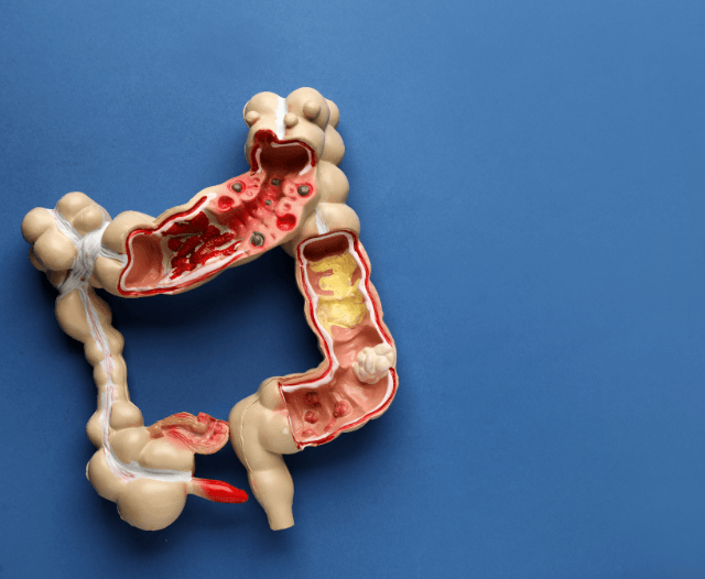
The small intestine is where the majority of nutrient absorption takes place.

It is divided into three sections: the duodenum, the jejunum, and the ileum.

Despite its essential role, the small intestine has historically been challenging to examine due to its length, intricate folds, and deep location within the body.


DBE_small_intestine


What Is Double Balloon Enteroscopy?

Double balloon enteroscopy (DBE) is a specialized endoscopic procedure that allows healthcare providers, typically gastroenterologists, to access and visualize the small intestine.

It is named for the use of two balloons – one on the endoscope and another on an overtube – which work together to advance and anchor the endoscope through the small intestine.

DBE is performed to diagnose and treat various small intestine conditions, such as Crohn’s disease, small bowel tumors, and unexplained gastrointestinal bleeding.

It is a minimally invasive procedure that offers significant advantages over traditional diagnostic methods, such as surgery.


When Is Double Balloon Enteroscopy Used?


Diagnostic Applications:


  1. Obscure Gastrointestinal Bleeding: DBE is especially valuable in diagnosing obscure gastrointestinal bleeding, where the source of bleeding is challenging to identify through other imaging techniques.
  2. Suspected Small Bowel Tumors: When small bowel tumors are suspected, DBE can provide a direct view of the lesions and collect tissue samples for biopsy.
  3. Evaluation of Crohn’s Disease: DBE is used to assess the extent and severity of Crohn’s disease in the small intestine, aiding in disease management and treatment planning.
  4. Unexplained Abdominal Pain: For individuals with chronic unexplained abdominal pain, DBE can help identify sources of discomfort within the small intestine.


Therapeutic Applications:

  1. Polyp and Tumor Removal: During DBE, gastroenterologists can remove small polyps and tumors from the small intestine, eliminating potential sources of future complications.
  2. Stricture Dilation: Strictures or narrowing in the small intestine can be dilated or stretched during DBE to relieve obstructions and improve the flow of food and liquids.
  3. Stent Placement: In cases of blockages or strictures, stents can be placed to maintain the patency of the small intestine and alleviate symptoms.


How Double Balloon Enteroscopy Works


The DBE procedure begins with the patient receiving sedation to ensure comfort and relaxation. Here is an overview of the key steps involved:

  1. Insertion: The endoscope, equipped with one balloon, is introduced through the mouth or rectum and guided into the small intestine.
  2. Balloon Inflation: The balloon on the endoscope is inflated to anchor it in place, while the overtube is advanced further into the small intestine.
  3. Advancement: The overtube is moved ahead, pushing the endoscope deeper into the small intestine.
  4. Visualization: The endoscope provides real-time images of the small intestine’s lining, allowing the gastroenterologist to closely examine the tissue and identify any abnormalities.
  5. Intervention: If polyps, tumors, or strictures are encountered, they can be treated during the procedure. This may involve removal, dilation, or stent placement, depending on the issue.
  6. Biopsy: Tissue samples (biopsies) can be collected for further analysis if necessary.


double_balloon_enteroscopy_procedure


What to Expect During a Double Balloon Enteroscopy


Before the procedure, you’ll receive specific instructions on preparation, which typically involves fasting and bowel preparation.

During the procedure:

  • You will be given sedation for comfort.
  • The procedure may take anywhere from 30 minutes to a few hours, depending on the extent of the examination and any interventions required.

After the procedure, you will be monitored until the sedation wears off. You may experience some mild abdominal discomfort, bloating, or cramping, which should subside within a day.


Recovery_after_dbe


Recovery and Aftercare


The recovery period following a DBE is relatively short.

Patients are typically able to resume their regular diet and activities the same day, although some may prefer to take it easy for a day or two.

The results of the procedure and any biopsies taken will be reviewed by your healthcare provider, who will discuss the findings and any necessary treatment or follow-up plans.


What are the risks of DBE?

Double balloon enteroscopy (DBE) is generally considered a safe and well-tolerated procedure.

However, like any medical procedure, it carries some inherent risks and potential complications.

These risks are typically low but need to be considered. It’s important for patients to be aware of these risks and discuss them with their healthcare provider before undergoing DBE.

Some of the potential risks and complications associated with DBE include:

  1. Bleeding: During DBE, biopsies may be taken, or polyps and tumors may be removed. In some cases, this can lead to bleeding. The majority of bleeding cases can be managed during the procedure, but there is a small risk of post-procedure bleeding, which might require further intervention.
  2. Perforation: Although rare, there is a risk of a small tear or perforation in the lining of the small intestine. Perforations can occur during the passage of the endoscope or during therapeutic interventions like polyp or stricture removal. Perforations are typically managed with immediate medical attention and, in some cases, surgical repair.
  3. Infection: Any time an invasive procedure is performed, there is a small risk of infection. To minimize this risk, DBE is performed in a sterile environment, and strict infection control protocols are followed.
  4. Reaction to Sedation: Sedative medications are administered to keep the patient comfortable and relaxed during the procedure. While these medications are generally safe, there is a small risk of an adverse reaction, such as an allergic reaction or breathing difficulties. It’s crucial for the healthcare team to monitor the patient’s condition during the procedure to address any potential complications promptly.
  5. Incomplete Examination: In some cases, it may not be possible to advance the endoscope through the entire small intestine. This can result in an incomplete examination and limit the ability to visualize certain areas of the small intestine. The reasons for an incomplete examination can vary, including anatomical factors or patient discomfort.
  6. Post-Procedure Discomfort: Patients may experience mild abdominal discomfort, bloating, or cramping following the procedure. This discomfort is usually short-lived and should resolve within a day.

It’s essential for patients to discuss these risks and any specific concerns with their healthcare provider before undergoing DBE.

Healthcare providers will assess the individual’s medical history and determine the appropriateness of the procedure based on their unique circumstances.


double_balloon_enteroscopy_infographic


Conclusion: Illuminating the Unseen with Double Balloon Enteroscopy


Double balloon enteroscopy is a remarkable advancement in the field of gastroenterology, providing healthcare providers with the means to explore and treat the small intestine.

With its diagnostic and therapeutic capabilities, DBE offers hope to individuals with small bowel conditions, including those with unexplained gastrointestinal bleeding, suspected tumors, or Crohn’s disease.

If you’re experiencing symptoms related to your small intestine or have been recommended for a DBE procedure, remember that this specialized tool can illuminate the unseen and pave the way for diagnosis, treatment, and ultimately, improved health and well-being.


Contact Dr. Deetlefs


Dr Eduan prides himself on his ability to help his patients to the best of his ability by embracing good listening skills, effective communication, compassion and knowledge and skill honed during years of private gastroenterology practice.

If you would like to book an appointment with a gastrointestinal (GI) specialist or would simply like more information on a particular GI topic, don’t hesitate to use our online booking form or call Dr. Deetlefs at 021 551 867. 

GIDoc Cape Town

Patient-focused GI treatments and procedures in Cape Town.

Monday-Friday 8AM-4PM.

Connect with Us

© Dr. Eduan Deetlefs, Registered Gastroenterologist, GI Doc Cape Town

Our website information is not intended or implied to be a substitute for professional medical advice, diagnosis or treatment. Please consult a doctor about your specific condition. Only a trained physician can determine an accurate diagnosis and proper treatment.

Gastroenterology Procedures Explained: A Comprehensive Guide

Gastroenterology Procedures Explained: A Comprehensive Guide


Gastroenterology is the medical specialty that focuses on the digestive system, encompassing the gastrointestinal (GI) tract, liver, pancreas, and gallbladder.

Gastroenterologists are specialists in diagnosing and treating a wide range of digestive disorders, from common conditions like acid reflux to complex diseases such as inflammatory bowel disease and liver cirrhosis.

To do this, they employ various procedures to examine and treat the GI system.

In this guide, we will explore these gastroenterology procedures in detail, shedding light on their purposes, how they are performed, and their significance in the field of digestive health.

Introduction to Gastroenterology Procedures


Gastroenterology procedures are invaluable tools that gastroenterologists use to diagnose, treat, and manage a wide array of gastrointestinal and hepatobiliary conditions.

These procedures provide a direct view of the internal structures of the digestive system, enabling accurate diagnosis and, in many cases, therapeutic interventions.

Diagnostic procedures are primarily used to assess the condition of the GI tract and adjacent organs, while therapeutic procedures involve the treatment of various digestive disorders.

Let’s delve into these procedures in detail, starting with diagnostic methods.

gastroenterology_capsule_endoscopy

Diagnostic Procedures


  • Upper Endoscopy (Esophagogastroduodenoscopy or EGD)
  • Colonoscopy
  • Flexible Sigmoidoscopy
  • Capsule Endoscopy
  • Endoscopic Ultrasound (EUS)
  • Imaging and Radiological Tests

Therapeutic Procedures


  • Polypectomy
  • Endoscopic Mucosal Resection (EMR)
  • Endoscopic Submucosal Dissection (ESD)
  • Esophageal Dilation
  • Gastrostomy and Jejunostomy Tubes


Advanced Gastroenterology Procedures


  • Endoscopic Retrograde Cholangiopancreatography (ERCP)
  • Percutaneous Endoscopic Gastrostomy (PEG)
  • Liver Biopsy


Diagnostic Procedures


Upper Endoscopy (Esophagogastroduodenoscopy or EGD)

Purpose: Upper endoscopy is a procedure that allows the gastroenterologist to examine the lining of the esophagus, stomach, and the upper part of the small intestine (duodenum). It is used to diagnose conditions like acid reflux, ulcers, gastritis, and Barrett’s esophagus.

Procedure: A thin, flexible tube with a light and camera at the tip is inserted through the mouth and guided down the throat. This provides real-time images of the upper GI tract, allowing the doctor to examine the tissues and take biopsies if necessary.

colonoscopy_procedure


Colonoscopy

Purpose: Colonoscopy is used to examine the large intestine (colon) and rectum. It is an essential tool for colorectal cancer screening and the diagnosis of conditions like inflammatory bowel disease and colorectal polyps.

Procedure: A long, flexible tube (colonoscope) with a camera is inserted through the rectum. It allows the doctor to visualize the colon’s inner lining and detect abnormalities. During the procedure, the gastroenterologist can also remove polyps or take biopsies.


Flexible Sigmoidoscopy

Purpose: This procedure focuses on examining the sigmoid colon, the last portion of the colon before the rectum. It is primarily used for colorectal cancer screening and the evaluation of lower GI tract symptoms.

Procedure: A flexible sigmoidoscope is inserted through the rectum to examine the sigmoid colon. It offers a limited view of the lower part of the colon, making it a valuable screening tool for certain conditions.


Capsule Endoscopy

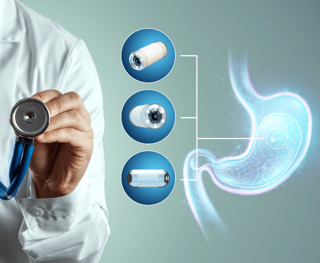
Purpose: Capsule endoscopy is a non-invasive method for imaging the small intestine. It is particularly useful for diagnosing conditions like Crohn’s disease and obscure gastrointestinal bleeding.

Procedure: Patients swallow a small, pill-sized capsule with a camera inside.

As the capsule moves through the digestive system, it captures images, which are transmitted to a receiver worn on the body. The images are later reviewed by the gastroenterologist.

 capsule_endoscopic_procedure

Endoscopic Ultrasound (EUS)

Purpose: EUS combines endoscopy and ultrasound to obtain detailed images of the digestive tract and nearby structures. It is often used for staging cancer and evaluating the pancreas and bile ducts.

Procedure: A thin, flexible tube is equipped with an ultrasound transducer and inserted through the mouth or rectum, depending on the area to be examined. EUS provides high-resolution images that help in diagnosing and staging diseases.


Imaging and Radiological Tests

In addition to endoscopic procedures, gastroenterologists also use various imaging and radiological tests to diagnose digestive and liver conditions:

  • Computed Tomography (CT) Scan: A non-invasive imaging technique used to assess the abdominal organs and surrounding structures.
  • Magnetic Resonance Imaging (MRI): MRI provides detailed images of the abdomen and is especially useful for evaluating the liver and bile ducts.
  • Ultrasound: Ultrasound is employed to visualize the liver, gallbladder, and blood vessels.
  • X-rays: X-ray studies with contrast agents, such as a barium swallow or enema, can reveal structural abnormalities in the GI tract.

polypectomy

Therapeutic Procedures


In addition to diagnosis, gastroenterologists perform a range of therapeutic procedures to manage and treat various gastrointestinal conditions:

Polypectomy

Purpose: Polyps are growths on the inner lining of the colon, and some can potentially develop into cancer. A polypectomy is performed during a colonoscopy to remove these polyps.

Procedure: A wire loop or snare is inserted through the colonoscope, allowing the gastroenterologist to remove polyps by cutting them from the colon wall.


Esophageal Dilation

Purpose: Esophageal dilation is performed to widen a narrowed or constricted esophagus, often due to conditions like strictures or Schatzki rings.

Procedure: A dilating device, such as a balloon or bougie, is used to gently stretch the esophagus, relieving symptoms like difficulty swallowing.


Gastrostomy and Jejunostomy Tubes

Purpose: Gastrostomy and jejunostomy tubes are placed in the stomach or jejunum (part of the small intestine) to provide enteral nutrition to patients who cannot eat or drink normally.

Procedure: A tube is inserted through the skin and into the stomach or jejunum to deliver liquid nutrition directly into the digestive tract.


Advanced Gastroenterology Procedures


Endoscopic Retrograde Cholangiopancreatography (ERCP)

Purpose: ERCP is a procedure to examine the bile ducts, pancreatic duct, and gallbladder. It is used for diagnosing and treating conditions like gallstones, pancreatitis, and bile duct obstructions.

Procedure: A flexible endoscope is passed through the mouth into the duodenum.

A contrast dye is injected into the ducts, and X-rays are taken to visualize any blockages or abnormalities. Therapeutic interventions, such as stone removal, can be performed during ERCP.


Percutaneous Endoscopic Gastrostomy (PEG)

Purpose: PEG is a method for placing a feeding tube directly into the stomach for long-term nutritional support, commonly used in individuals who cannot eat or drink normally.

Procedure: A tube is inserted through the abdominal wall and into the stomach, allowing for the administration of nutrition and medications.


Liver Biopsy

Purpose: A liver biopsy is performed to obtain a small sample of liver tissue for examination, aiding in the diagnosis and staging of liver diseases.

Procedure: A needle is inserted through the skin into the liver, and a small tissue sample is collected for analysis.

Conclusion: The Evolving Landscape of Gastroenterology Procedures


Gastroenterology procedures have revolutionized the diagnosis and treatment of gastrointestinal and hepatobiliary conditions.

They provide gastroenterologists with the tools to not only understand the inner workings of the digestive system but also to intervene and treat various disorders effectively.

As technology and research continue to advance, the field of gastroenterology will only become more precise, patient-friendly, and outcomes-focused.

This evolution offers hope to individuals dealing with digestive diseases, as it promises more accurate diagnoses, less invasive treatments, and improved overall care.

For individuals with gastrointestinal symptoms or those at risk of digestive conditions, seeking the expertise of a gastroenterologist and considering these procedures, when necessary, can lead to better outcomes and a higher quality of life.

Gastroenterology procedures are not just medical tools; they are pathways to understanding, treatment, and digestive well-being.


gastroenterology_procedures_infographic


Contact Dr. Deetlefs


Dr Eduan prides himself on his ability to help his patients to the best of his ability by embracing good listening skills, effective communication, compassion and knowledge and skill honed during years of private gastroenterology practice.

If you would like to book an appointment with a gastrointestinal (GI) specialist or would simply like more information on a particular GI topic, don’t hesitate to use our online booking form or call Dr. Deetlefs at 021 551 867.

GIDoc Cape Town

Patient-focused GI treatments and procedures in Cape Town.

Monday-Friday 8AM-4PM.

Connect with Us

© Dr. Eduan Deetlefs, Registered Gastroenterologist, GI Doc Cape Town

Our website information is not intended or implied to be a substitute for professional medical advice, diagnosis or treatment. Please consult a doctor about your specific condition. Only a trained physician can determine an accurate diagnosis and proper treatment.

Gastrointestinal Health: Nurturing Your Gut for Well-Being

Gastrointestinal Health: Nurturing Your Gut for Well-Being


Gastrointestinal health, often referred to as gut health, is a fundamental pillar of overall well-being.

The digestive system, with its intricate mechanisms and microbiome, plays a crucial role in maintaining our health.

In this comprehensive guide, we’ll explore the intricacies of gastrointestinal health, understand its significance, and discuss practical steps to nurture and optimize your gut for a healthier and happier life.

The Marvel of the Gastrointestinal System

The gastrointestinal system, also known as the digestive system, is a marvel of engineering.

It begins at the mouth, where digestion commences, and extends through the esophagus, stomach, small intestine, large intestine, and finally, the rectum and anus.

This system’s primary function is to process food and extract essential nutrients, vitamins, and minerals while eliminating waste.

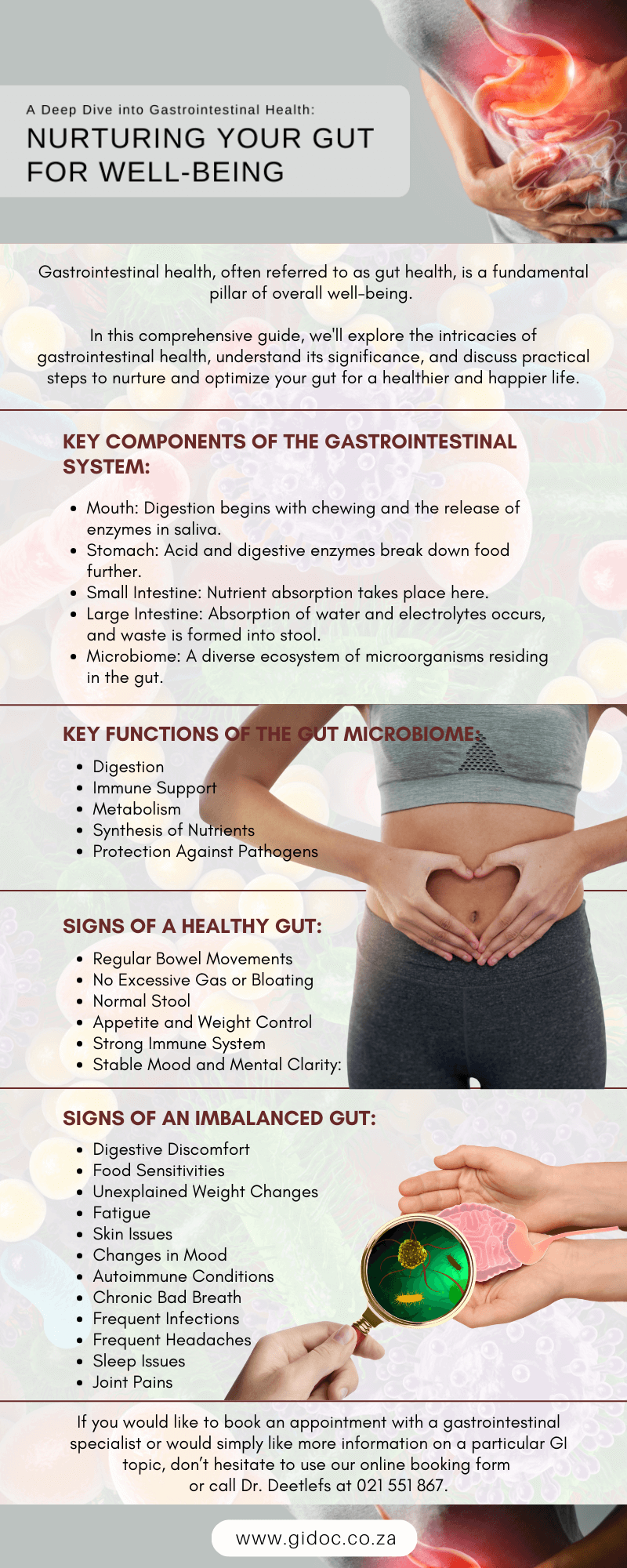
Key Components of the Gastrointestinal System:

Mouth: Digestion begins with chewing and the release of enzymes in saliva.

Stomach: Acid and digestive enzymes break down food further.

Small Intestine: Nutrient absorption takes place here.

Large Intestine: Absorption of water and electrolytes occurs, and waste is formed into stool.

Microbiome: A diverse ecosystem of microorganisms residing in the gut.

gut_microbiome


Understanding the Gut Microbiome

The gut microbiome is an ecosystem of trillions of microorganisms, including bacteria, viruses, fungi, and other microbes.

It’s like a bustling city of microorganisms that play a critical role in digestion, metabolism, and even immune system function.

The composition and diversity of your gut microbiome can profoundly impact your health.

Key Functions of the Gut Microbiome:

Digestion: Gut bacteria assist in breaking down complex carbohydrates and fibers that the human body cannot digest on its own.

Immune Support: A healthy microbiome helps the immune system function optimally.

Metabolism: The gut microbiome can influence how the body stores fat and regulates blood sugar.

Synthesis of Nutrients: Some gut bacteria produce essential vitamins and nutrients.

Protection Against Pathogens: A diverse microbiome can help prevent the overgrowth of harmful bacteria.

Signs of a Healthy Gut

A well-functioning gastrointestinal system exhibits several signs of good health:

Regular Bowel Movements: Consistent, comfortable, and regular bowel movements are a hallmark of a healthy gut.

No Excessive Gas or Bloating: Occasional gas is normal, but excessive or chronic bloating and gas may indicate an imbalance.

Normal Stool: Healthy stool should be well-formed and easy to pass.

Appetite and Weight Control: A healthy gut helps regulate appetite and supports a balanced weight.

Strong Immune System: A robust immune response is a sign of a well-supported immune system.

Stable Mood and Mental Clarity: Emerging research suggests a connection between the gut and mental health.

Diagnosing an Unhealthy Gut:

Diagnosing an unhealthy gut or gut dysbiosis typically involves a combination of medical history, physical examination, and, in some cases, laboratory tests.

Here’s how healthcare providers diagnose an unhealthy gut:

  1. Medical History: A thorough medical history is an essential part of the diagnostic process. During this conversation, your healthcare provider will ask about your gastrointestinal symptoms, including diarrhea, constipation, bloating, abdominal pain, and changes in bowel habits.
  1. Physical Examination: A physical examination may be performed to assess your overall health and identify any signs of gastrointestinal distress.
  2. Stool Analysis: Stool tests can provide valuable insights into the composition of your gut microbiome and the presence of any imbalances.
  3. Blood Tests: Blood tests can be conducted to assess markers of inflammation and nutrient levels, which can indicate gut-related issues.
  4. Endoscopic Procedures: In some cases, your healthcare provider may recommend an endoscopy or colonoscopy.

diagnsoing_unhealthy_gut

The Connection Between Gut Health and Overall Well-Being

Gastrointestinal health is intricately linked to the body’s overall well-being. Recent scientific research has unveiled the gut-brain axis, a communication network that connects the gut and the brain.

What are some common signs of an imbalanced gut?

An imbalanced gut, often referred to as gut dysbiosis, can manifest in various ways. While not all signs may be present, here are some common symptoms and signs that may indicate an imbalance in your gut microbiome:

Digestive Discomfort: One of the most noticeable signs is persistent digestive discomfort, which can include bloating, gas, abdominal pain, cramping, and irregular bowel movements (diarrhea or constipation).

Food Sensitivities: An imbalanced gut can lead to increased sensitivity to certain foods, resulting in digestive symptoms like bloating, diarrhea, or stomach cramps after consumption.

Unexplained Weight Changes: Significant and unexplained weight loss or gain can be linked to an imbalanced gut, as the microbiome plays a role in nutrient absorption and metabolism.

Fatigue: An imbalanced gut may affect nutrient absorption, potentially leading to anemia or nutrient deficiencies, which can cause fatigue.

Skin Issues: Skin conditions such as acne, eczema, or rosacea can be associated with gut dysbiosis, as the gut and skin are interconnected.

Changes in Mood: Emerging research suggests a link between gut health and mental well-being. Anxiety, depression, and mood swings can sometimes be connected to gut imbalances.

Autoimmune Conditions: Some autoimmune conditions have been linked to imbalances in the gut, as gut health is closely related to the immune system.

Chronic Bad Breath: An imbalanced gut can lead to bad breath that doesn’t improve with oral hygiene practices.

Frequent Infections: A weakened gut can make you more susceptible to infections as a balanced gut microbiome plays a significant role in the immune system.

Frequent Headaches: Recurring headaches or migraines can sometimes be related to an imbalanced gut.

Sleep Issues: An imbalanced gut can affect sleep patterns, causing difficulties with both falling asleep and staying asleep.

Joint Pain: In some cases, joint pain and conditions like rheumatoid arthritis have been associated with gut dysbiosis.

Chronic Diarrhea or Constipation: An imbalance can lead to persistent changes in bowel habits, including frequent diarrhea or long-term constipation.

Skin Rashes or Irritations: Conditions like psoriasis, dermatitis, or hives may be related to gut health.

Autoimmune Disorders: Some autoimmune disorders, like celiac disease, are closely linked to the gut, with the immune system mistakenly attacking the body’s own tissues.

It’s important to note that these symptoms can have various causes, and experiencing one or more of them doesn’t necessarily mean you have gut dysbiosis.

unhealhty_gut_symptoms

Nurturing Your Gut for Better Health

Caring for your gastrointestinal health is a proactive step towards improved overall well-being. Here are some practical strategies to optimize your gut health:

Eat a Diverse Diet:
A varied diet rich in fiber, fruits, vegetables, and whole grains supports gut health by promoting a diverse microbiome.

Include Fermented Foods:
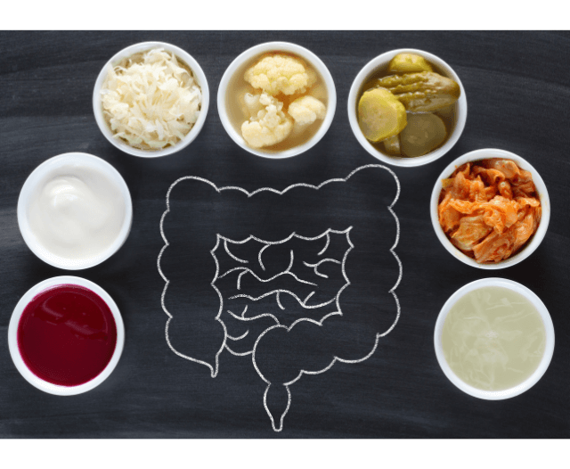
Yogurt, kefir, sauerkraut, and kimchi contain beneficial probiotics that contribute to gut health.

Stay Hydrated:
Adequate water intake is essential for digestive health and the absorption of nutrients.

Manage Stress:
Practicing stress-reduction techniques such as meditation and yoga can help maintain a healthy gut-brain axis.

Avoid Excessive Antibiotic Use:
Overuse of antibiotics can disrupt the gut microbiome. Use them only when necessary.

Get Regular Exercise:
Physical activity supports gastrointestinal motility and overall health.

Limit Processed Foods:
Processed foods often contain additives that can negatively affect the gut. Opt for whole, unprocessed foods.

Sleep Well:
Quality sleep is essential for a healthy gut and overall well-being.

Be Mindful of Food Sensitivities:
Identify and manage any food sensitivities or allergies that may disrupt your gut.

Stay Informed: 
Keep up with the latest research on gut health to make informed choices about your diet and lifestyle.

When to Seek Medical Advice

While many digestive issues can be managed with lifestyle changes, it’s crucial to seek medical advice if you experience persistent or severe gastrointestinal symptoms, unexplained weight loss, or if you have a family history of gastrointestinal conditions.

A healthcare provider can diagnose and provide guidance on the management of conditions like irritable bowel syndrome (IBS), Crohn’s disease, and ulcerative colitis.

gastrointestinal_health_gut

Contact Dr. Deetlefs

Dr Eduan prides himself on his ability to help his patients to the best of his ability by embracing good listening skills, effective communication, compassion and knowledge and skill honed during years of private gastroenterology practice.

If you would like to book an appointment with a gastrointestinal (GI) specialist or would simply like more information on a particular GI topic, don’t hesitate to use our online booking form or call Dr. Deetlefs at 021 551 867.

GIDoc Cape Town

Patient-focused GI treatments and procedures in Cape Town.

Monday-Friday 8AM-4PM.

Connect with Us

© Dr. Eduan Deetlefs, Registered Gastroenterologist, GI Doc Cape Town

Our website information is not intended or implied to be a substitute for professional medical advice, diagnosis or treatment. Please consult a doctor about your specific condition. Only a trained physician can determine an accurate diagnosis and proper treatment.

Exploring ERCP: A Comprehensive Guide

Exploring ERCP: A Comprehensive Guide

This versatile and powerful procedure has revolutionized the field of gastroenterology, allowing healthcare professionals to diagnose and treat a wide range of conditions affecting the bile ducts, pancreas, and gallbladder.

In this comprehensive guide, we will delve into the world of ERCP, exploring its uses, benefits, procedure, potential risks, and much more.

Understanding ERCP: What Is It?

Endoscopic Retrograde Cholangiopancreatography (ERCP) is a specialized medical procedure that combines endoscopy and fluoroscopy to diagnose and treat conditions in the bile ducts, pancreas, and gallbladder.

It provides detailed images and, if necessary, allows for the performance of therapeutic interventions. ERCP is typically performed by a gastroenterologist, a specialist in digestive diseases.

The Key Components of ERCP

Endoscope: An endoscope is a flexible, tube-like instrument equipped with a light source and a camera. It is inserted through the mouth, down the esophagus, and into the stomach and duodenum (the first part of the small intestine).

Fluoroscopy: Fluoroscopy is a real-time X-ray imaging technique that provides dynamic images of the bile ducts and pancreatic duct.

Contrast Medium: A contrast medium, often a special dye, is injected into the ducts to make them visible on X-ray images.

ercp_procedure

When Is ERCP Used?

ERCP is employed for various diagnostic and therapeutic purposes, primarily related to the digestive system. Some common indications for ERCP include:

  1. Diagnosing and Treating Gallstones:

ERCP can identify gallstones trapped in the bile ducts, causing symptoms like jaundice, abdominal pain, and pancreatitis (inflammation of the pancreas). In such cases, the stones can be removed during the procedure.

  1. Diagnosing and Managing Biliary and Pancreatic Tumors:

ERCP can help in evaluating and diagnosing tumors or strictures (narrowing) in the bile ducts or pancreatic duct. It can also assist in placing stents to relieve blockages caused by these conditions.

  1. Treating Pancreatitis:
    In some cases of acute pancreatitis, ERCP may be performed to remove obstructions or perform other therapeutic procedures.
  1. Draining Fluid Collections:

ERCP can be used to drain fluid collections (pseudocysts) in the pancreas or bile ducts.

  1. Evaluating Chronic Abdominal Pain:
    When the cause of chronic abdominal pain is unclear, ERCP can be used as a diagnostic tool to explore the pancreas and bile ducts.
  1. Roux-en-Y Gastric Bypass Procedures:
    In patients who have previously undergone gastric bypass surgery, ERCP can be challenging. However, specialized techniques, such as laparoscopy-assisted or double-balloon-assisted ERCP, have been developed to access the desired areas.

ercp_balloon_enteroscopy

The ERCP Procedure: What to Expect

Before undergoing ERCP, patients are typically given instructions about fasting to ensure an empty stomach. The procedure is performed in a hospital or outpatient endoscopy center and generally follows these steps:

  1. Preparation:

Patients may receive a sedative or anesthesia to relax and minimize discomfort during the procedure. The throat is also numbed with a local anesthetic to ease the insertion of the endoscope.

  1. Insertion of the Endoscope:

The gastroenterologist carefully inserts the endoscope through the mouth and into the esophagus, stomach, and duodenum.

  1. Locating the Papilla:

The papilla of Vater, a small nipple-like structure where the common bile duct and pancreatic duct drain into the duodenum, is located using the endoscope.

  1. Contrast Injection:

A contrast medium (dye) is injected into the ducts to make them visible on X-ray images. Fluoroscopy is used to monitor the movement of the contrast medium.

  1. Imaging and Diagnosis:

X-ray images are captured in real-time to evaluate the structure and function of the bile ducts and pancreatic duct. If any abnormalities are detected, they can be further examined or treated during the same procedure.

  1. Therapeutic Procedures:

ERCP allows for various therapeutic interventions, such as removing gallstones, placing stents, widening strictures, or draining fluid collections. These procedures can alleviate symptoms and improve overall health.

  1. Completion:

Once the necessary diagnostic or therapeutic steps are completed, the endoscope is carefully removed.
The duration of an ERCP can vary but typically takes between 30 minutes to an hour. After the procedure, patients are monitored in a recovery area until the sedation wears off, and it is safe to be discharged.

Potential Risks and Complications of ERCP

While ERCP is generally considered a safe and effective procedure, like any medical intervention, it carries some risks and potential complications.

These can include:

  1. Pancreatitis: This is one of the most common complications of ERCP. Pancreatitis is inflammation of the pancreas and can range from mild to severe. It can cause abdominal pain, nausea, vomiting, and in severe cases, may require hospitalization. The risk of pancreatitis following ERCP varies but is generally around 5% to 10%. It is more common when therapeutic interventions, such as sphincterotomy (cutting of the sphincter), are performed during the procedure.
  2. Infection: Infections can occur in rare cases, either in the bile ducts or the pancreas. Bacterial infection can lead to symptoms such as fever, chills, and abdominal pain. Infection risk is minimized by following strict sterile techniques during the procedure.
  3. Bleeding: Although uncommon, bleeding can occur during or after ERCP, particularly if therapeutic procedures like sphincterotomy or stone removal are performed. Minor bleeding may resolve on its own, but significant bleeding may require additional interventions or surgery.
  4. Perforation: Perforation, or a tear in the digestive tract, is a rare but serious complication. It can lead to abdominal pain, infection, and the leakage of digestive contents into the abdominal cavity. Emergency surgery is usually necessary to repair a perforation.
  5. Allergic Reaction: Some patients may experience an allergic reaction to the contrast medium used during ERCP. Allergic reactions can range from mild skin rashes to more severe symptoms like difficulty breathing or anaphylaxis. The risk of a severe allergic reaction is relatively low.
  6. Complications Related to Anesthesia: Sedatives and anesthesia used during ERCP carry inherent risks, including respiratory depression, reduced oxygen levels, and reactions to medications. These risks are typically minimized by careful monitoring during the procedure.
  7. Other Risks: There is also a slight risk of complications such as blood clots, heart problems, or adverse reactions to medications used during ERCP.

It’s important to note that while these risks are associated with ERCP, the procedure is generally considered safe and effective when performed by experienced gastroenterologists or endoscopists.

 ercp_after_procedure

Recovery and Aftercare

After an ERCP, patients are typically monitored for a brief period in a recovery area until they fully wake up from the sedation. It’s essential to arrange for a friend or family member to drive them home, as the sedatives can impair driving ability.

Patients may experience mild discomfort, bloating, or a sore throat for a day or two after the procedure, but these symptoms generally resolve on their own. If any complications or severe symptoms arise, such as persistent abdominal pain, fever, chills, or heavy bleeding, patients should contact their healthcare provider immediately.

Conclusion: The Power of ERCP in Gastroenterology

Endoscopic Retrograde Cholangiopancreatography (ERCP) is a powerful tool in the field of gastroenterology, offering both diagnostic and therapeutic capabilities.

By combining endoscopy and fluoroscopy, this procedure allows healthcare professionals to explore and treat conditions affecting the bile ducts, pancreas, and gallbladder with minimal invasiveness.

While ERCP can carry some risks and potential complications, it has greatly improved the accuracy of diagnoses and the effectiveness of treatments for a wide range of gastrointestinal conditions.

When performed by skilled and experienced gastroenterologists, ERCP can be a life-changing procedure that provides relief and improved quality of life for patients.

ercp_risks_benefits_infographic

Contact Dr. Deetlefs

Dr Eduan prides himself on his ability to help his patients to the best of his ability by embracing good listening skills, effective communication, compassion and knowledge and skill honed during years of private gastroenterology practice.

If you would like to book an appointment with a gastrointestinal (GI) specialist or would simply like more information on a particular GI topic, don’t hesitate to use our online booking form or call Dr. Deetlefs at 021 551 867.

GIDoc Cape Town

Patient-focused GI treatments and procedures in Cape Town.

Monday-Friday 8AM-4PM.

Connect with Us

© Dr. Eduan Deetlefs, Registered Gastroenterologist, GI Doc Cape Town

Our website information is not intended or implied to be a substitute for professional medical advice, diagnosis or treatment. Please consult a doctor about your specific condition. Only a trained physician can determine an accurate diagnosis and proper treatment.

Exploring the Future of Diagnosis: Capsule Endoscopy

Exploring the Future of Diagnosis: Capsule Endoscopy

Overview

In the realm of medical diagnostics, innovation has consistently pushed the boundaries of what is possible.

Capsule endoscopy, a relatively recent addition to the medical toolkit, exemplifies this trend.

This remarkable technology has revolutionized the field of gastroenterology, allowing healthcare professionals to explore the inner workings of the digestive tract in a minimally invasive and patient-friendly manner.

In this comprehensive guide, we will journey into the world of capsule endoscopy, delving into its history, mechanics, applications and benefits.


What does it look like?


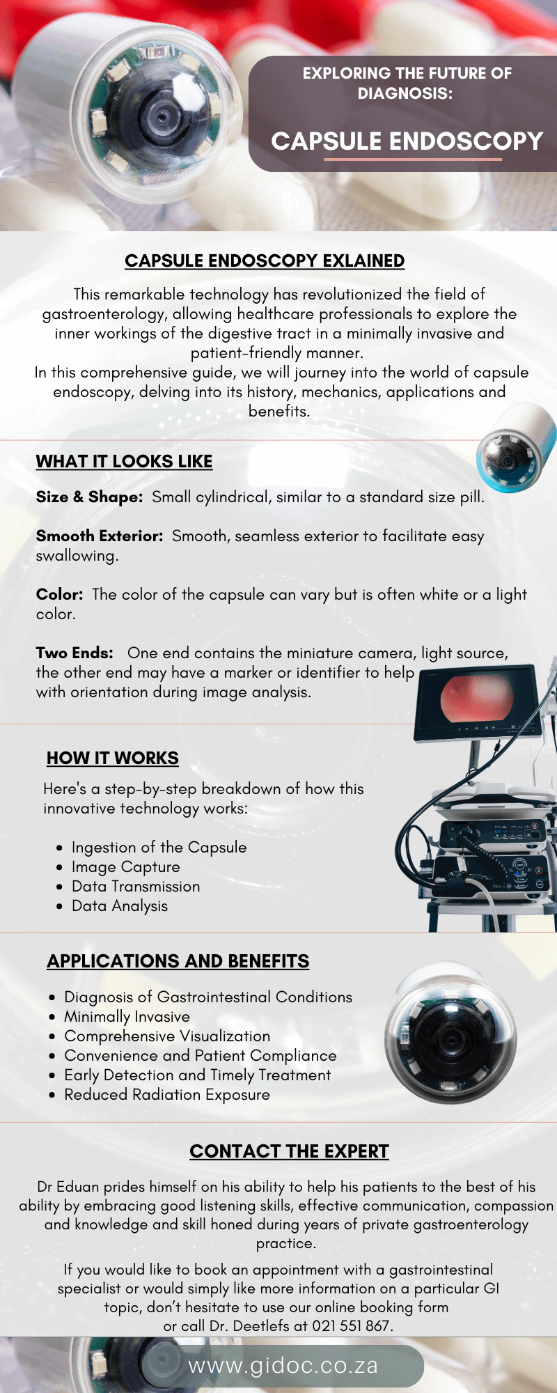
A capsule endoscopy, often referred to as a “pill camera,” is a small, ingestible medical device that is about the size and shape of a large vitamin pill.

Its appearance can vary slightly depending on the manufacturer, but here are some common characteristics:

Size and Shape: A typical capsule endoscopy capsule is small and cylindrical, similar in size to a standard pill. It is designed to be easy to swallow, typically measuring around 26 mm in length and 11 mm in diameter.

Smooth Exterior: The capsule has a smooth, seamless exterior to facilitate easy swallowing and safe passage through the gastrointestinal (GI) tract. It’s typically made of medical-grade materials that are safe for ingestion.

Color: The color of the capsule can vary but is often white or a light color. This helps with visibility if the capsule is ever seen in the patient’s stool.

Two Ends: Capsule endoscopy capsules typically have two ends. One end contains the miniature camera, light source, and other imaging components. The other end may have a marker or identifier to help with orientation during image analysis.

It’s important to note that while the appearance of the capsule itself is relatively simple, the technology it contains is highly sophisticated.

The miniature camera and imaging components inside the capsule are capable of capturing high-quality images of the gastrointestinal tract, providing valuable diagnostic information to healthcare providers. 

capsul;e_endoscopy_benefits


The Journey Begins: A Brief History of Capsule Endoscopy


Before we delve into the intricacies of capsule endoscopy, it’s essential to understand its origins and evolution.

The story of capsule endoscopy traces back to the late 20th century when medical researchers began seeking alternatives to traditional endoscopy procedures, which often involved discomfort and anesthesia.


1. Development of the Capsule Camera

The concept of a miniature camera that could travel through the gastrointestinal (GI) tract began to take shape in the late 1990s.

Researchers envisioned a capsule-sized device equipped with a tiny camera, light source, and transmission system that could capture images as it passed through the digestive system.

This idea laid the foundation for what would become the capsule endoscope.


2. The PillCam Era

In the early 2000s, Israeli company Given Imaging introduced the “PillCam.”

This small, swallowable capsule endoscope, which resembled a pill, marked a significant breakthrough in medical imaging.

The PillCam’s wireless technology allowed for the transmission of real-time images as it traversed the GI tract, providing detailed views of the esophagus, stomach, small intestine, and even parts of the colon.

The PillCam SB, designed for small bowel examination, received FDA approval in 2001, paving the way for capsule endoscopy to become a viable diagnostic tool.


The Capsule Unveiled: How Capsule Endoscopy Works


Capsule endoscopy is a procedure that employs a small, ingestible capsule containing a miniaturized camera, light source, battery, and wireless transmitter. Here’s a step-by-step breakdown of how this innovative technology works:


1. Ingestion of the Capsule

The procedure begins with the patient swallowing the capsule, which is about the size of a large vitamin pill. It is attached to a thin, flexible cord that transmits the images to a data recorder worn by the patient. The capsule’s journey through the GI tract is entirely passive, driven by peristalsis – the natural muscular contractions of the digestive system.


2. Image Capture

As the capsule progresses through the digestive tract, the camera continuously captures high-quality images of the lining of the GI tract. These images provide detailed views of the mucosa, allowing for the detection of abnormalities such as ulcers, polyps, bleeding, inflammation, and tumors.


3. Data Transmission

The images are transmitted wirelessly from the capsule to the data recorder, which the patient wears on their body. This recorder is about the size of a small smartphone and is connected to adhesive patches on the patient’s abdomen. It stores the images and allows for continuous monitoring of the procedure.


4. Data Analysis

After the capsule endoscopy is completed, typically within eight hours, the patient returns the data recorder to the healthcare provider. The recorded images are then downloaded to a computer for analysis. Advanced software assists in reviewing and interpreting the images, enabling the healthcare provider to make a diagnosis.

capsule_endoscopy_exploring_the_future


Capsule Endoscopy: Applications and Benefits


Capsule endoscopy has rapidly gained popularity due to its numerous applications and several significant advantages over traditional endoscopy methods. Let’s explore some of the key applications and benefits:

1. Diagnosis of Gastrointestinal Conditions

Capsule endoscopy is primarily used to diagnose a wide range of gastrointestinal conditions, including:

  • Small Bowel Diseases: It is particularly effective in detecting small bowel diseases like Crohn’s disease, celiac disease, and small bowel tumors.
  • Obscure Gastrointestinal Bleeding: Capsule endoscopy is invaluable in identifying the source of obscure gastrointestinal bleeding when other diagnostic methods fail.
  • Polyp and Tumor Detection: It can detect polyps and tumors in the esophagus, stomach, and small intestine.

2. Minimally Invasive

One of the most significant advantages of capsule endoscopy is its minimally invasive nature.
Unlike traditional endoscopy procedures, which require the insertion of a long, flexible tube through the mouth or rectum, capsule endoscopy involves no discomfort, sedation, or anesthesia.
This makes it more patient-friendly and reduces the risk of complications.


3. Comprehensive Visualization

Capsule endoscopy provides comprehensive visualization of the entire small intestine, a part of the GI tract that is challenging to reach with traditional endoscopy techniques.
This allows for a more thorough assessment of the digestive system.

  1. Convenience and Patient Compliance
    Patients often find capsule endoscopy more convenient and less intimidating than traditional endoscopy. The ability to swallow a small capsule eliminates the need for hospital visits and anesthesia, making it more appealing to many individuals.

5. Early Detection and Timely Treatment

The high-quality images generated by capsule endoscopy enable the early detection of gastrointestinal conditions. Early diagnosis often leads to timely treatment, improving patient outcomes and reducing the risk of complications.

6. Reduced Radiation Exposure

Capsule endoscopy does not involve the use of ionizing radiation, making it a safer option for certain patient populations, including pregnant women and individuals who require repeated diagnostic procedures. 


Conclusion


Capsule endoscopy represents a remarkable leap forward in the field of gastroenterology.

Its minimally invasive nature, comprehensive visualization capabilities, and patient-friendly approach have made it a valuable tool for diagnosing a wide range of gastrointestinal conditions.

As technology continues to evolve, the future of capsule endoscopy holds the promise of even more advanced diagnostic capabilities, ultimately leading to improved patient care and outcomes.

With each capsule that embarks on its journey through the digestive tract, the world of medical diagnostics moves one step closer to unlocking the secrets of the human body.

Dr Eduan prides himself on his ability to help his patients to the best of his ability by embracing good listening skills, effective communication, compassion and knowledge and skill honed during years of private gastroenterology practice.

If you would like to book an appointment with a gastrointestinal (GI) specialist or would simply like more information on a particular GI topic, don’t hesitate to use our online booking form or call Dr. Deetlefs at 021 551 867.


GIDoc Cape Town

Patient-focused GI treatments and procedures in Cape Town.

Monday-Friday 8AM-4PM.

Connect with Us

© Dr. Eduan Deetlefs, Registered Gastroenterologist, GI Doc Cape Town

Our website information is not intended or implied to be a substitute for professional medical advice, diagnosis or treatment. Please consult a doctor about your specific condition. Only a trained physician can determine an accurate diagnosis and proper treatment.